“. . . according to the Yoga Sutra (3.1), the term [Bandha] refers to the ‘binding’ of consciousness to a particular object or locus (desha), which is the very essence of concentration.”
Georg Feuerstein


Shoulder Biomechanics, Part III: The Supraspinatus Muscle

Hello Friends,

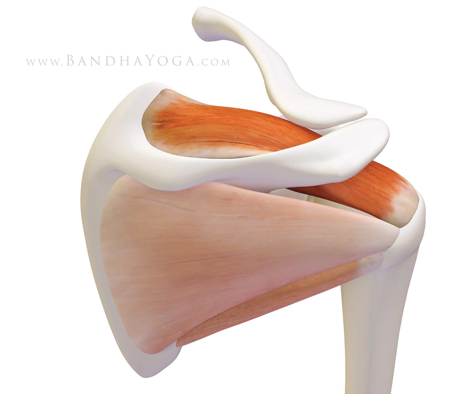
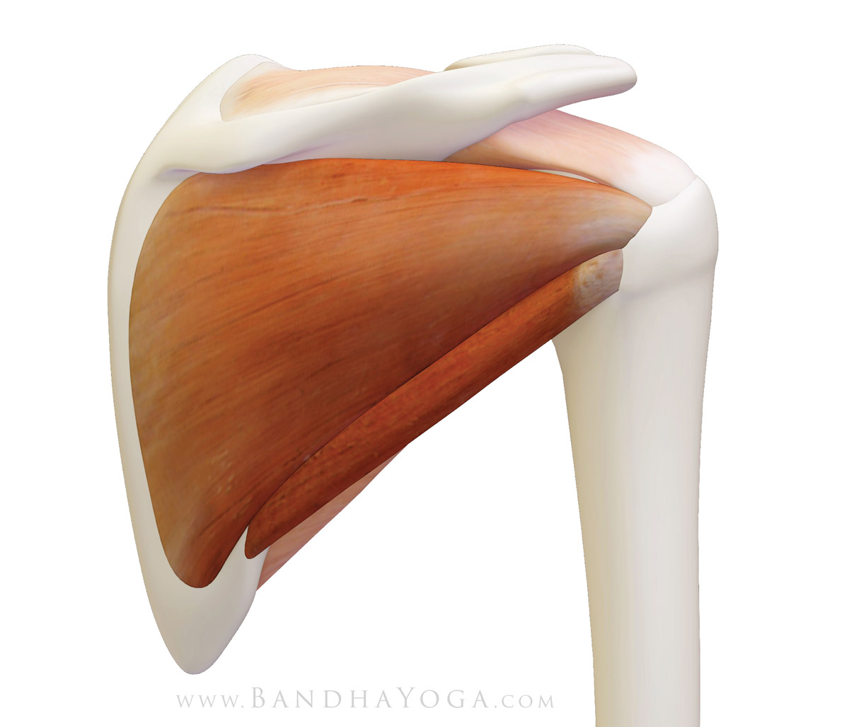
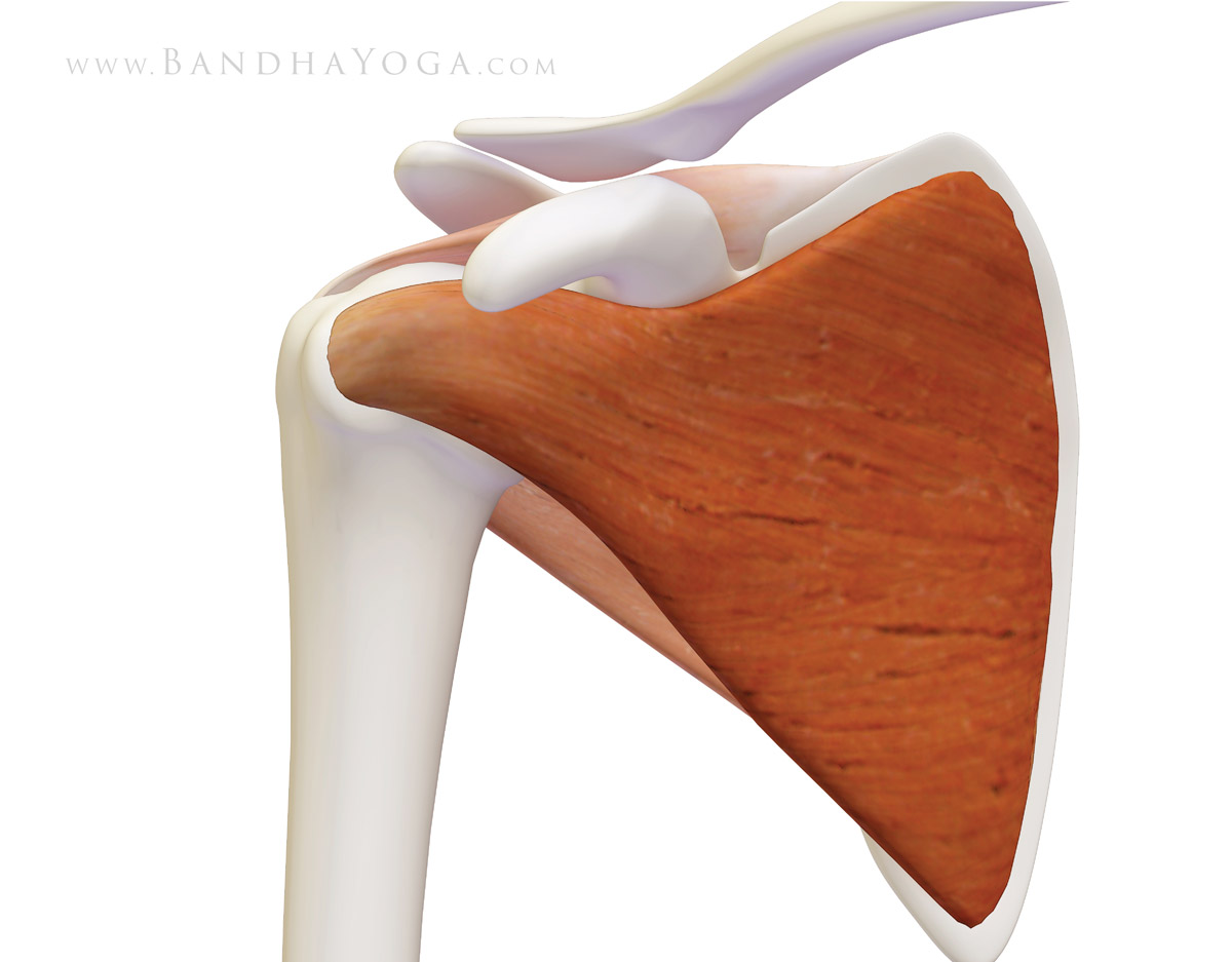
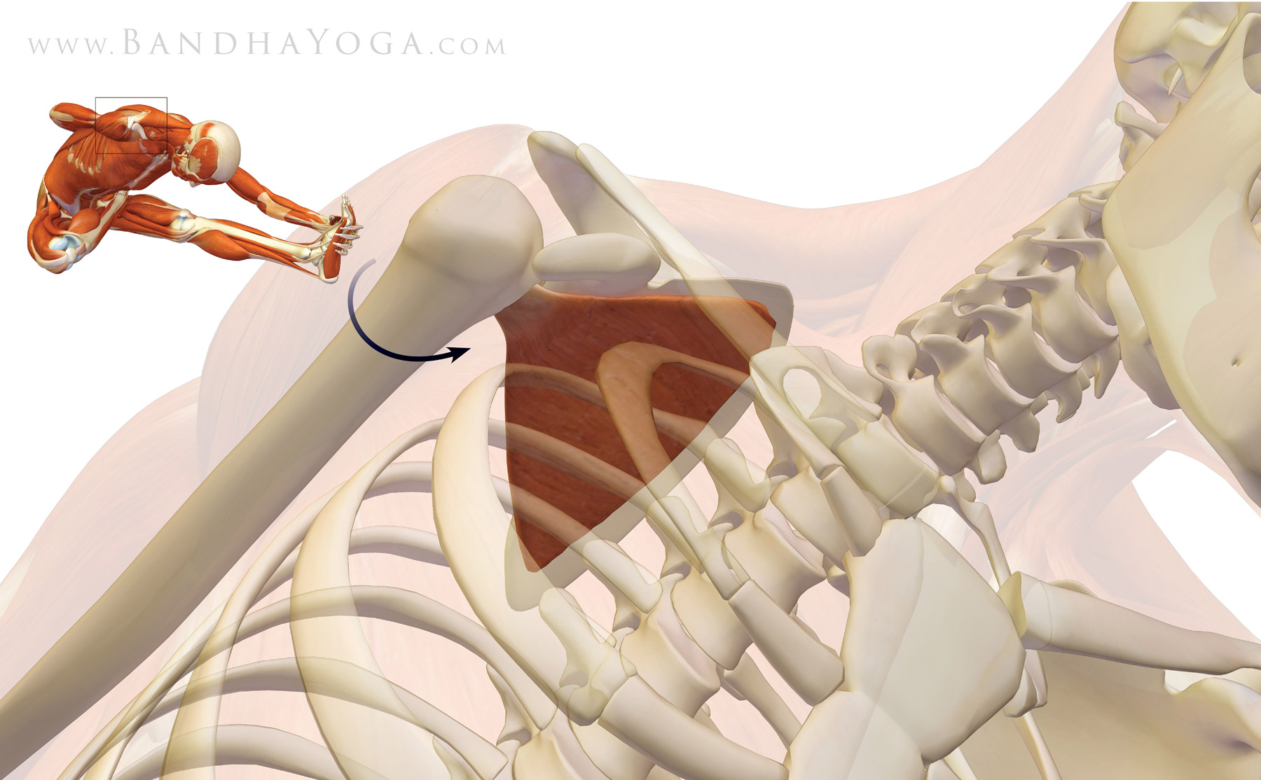
Let’s cap off the muscles of your rotator cuff with the supraspinatus. This muscle originates in a trough-like area above the scapular spine, hence its name supra, which means “above”. The supraspinatus then inserts onto the greater tuberosity just behind where the long head of the biceps enters the shoulder (figure 1).

(We’ve covered the subscapularis, infraspinatus and teres minor muscles along with some key biomechanical points about each muscle—click to review.)


Figure 1: The supraspinatus muscle of the rotator cuff (with the infraspinatus and teres minor faded).


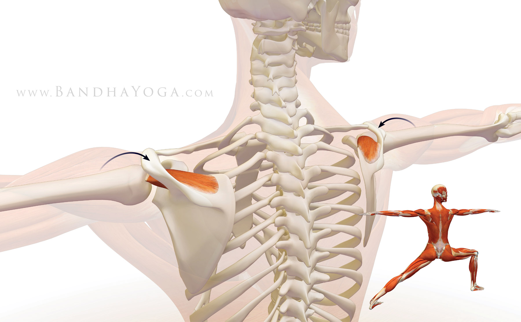
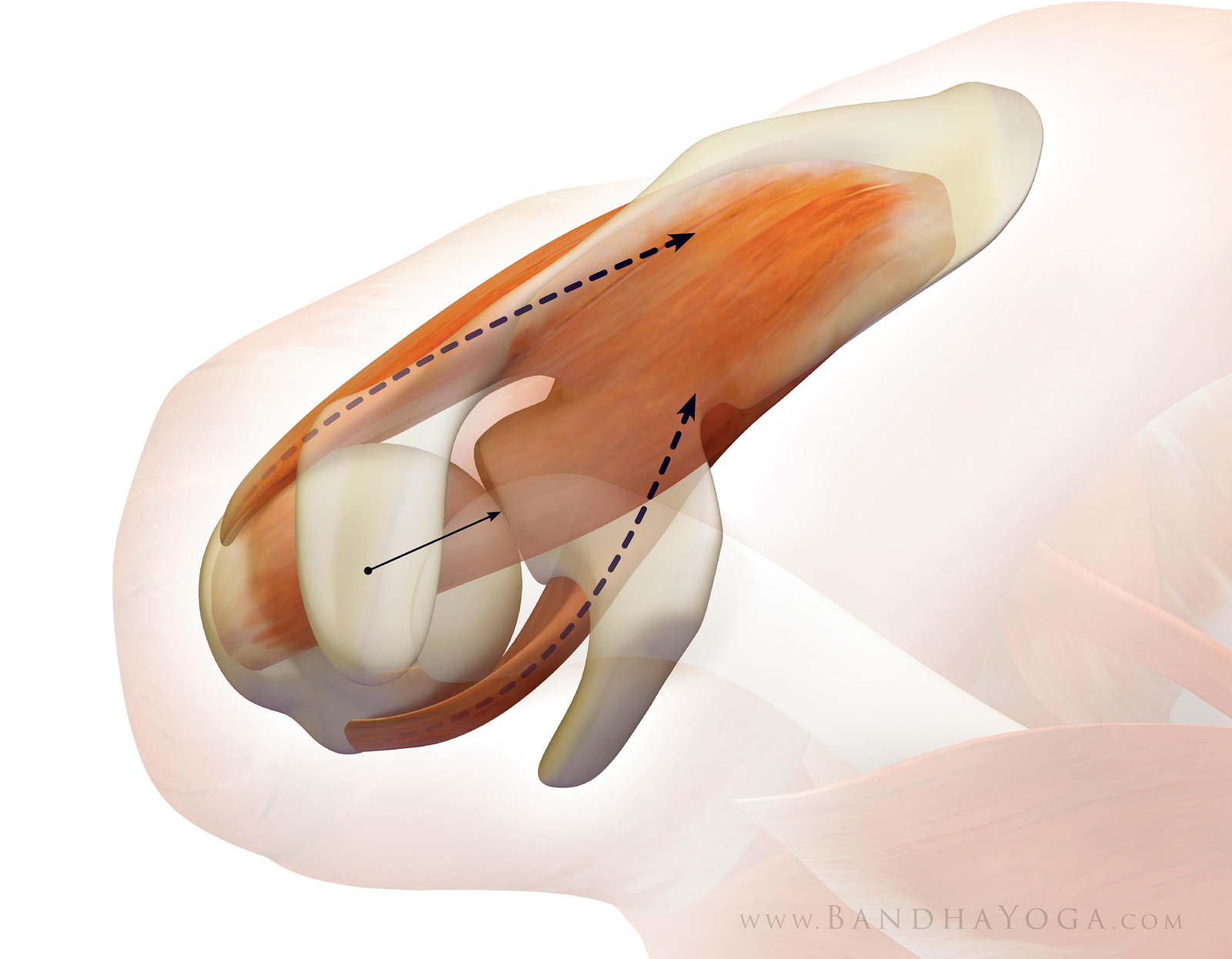
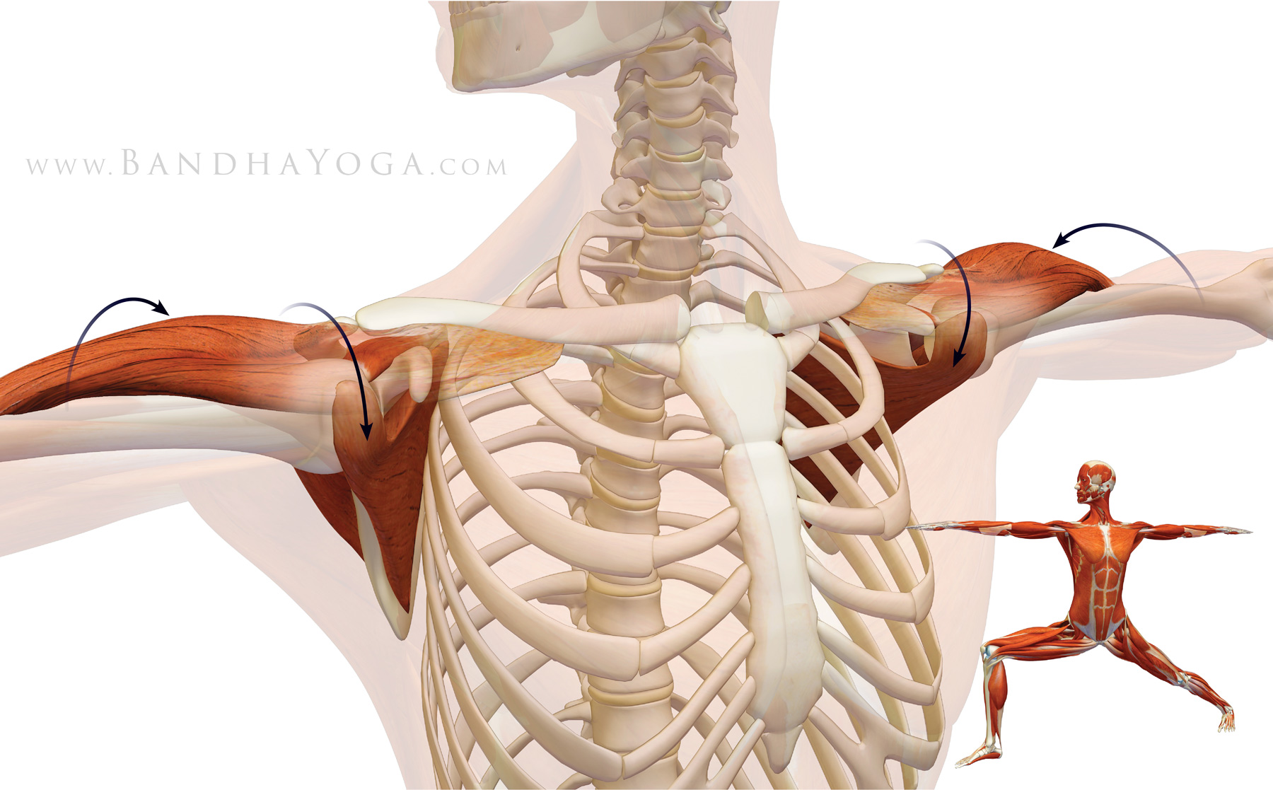
Contracting the supraspinatus abducts the humerus at the glenoid socket (takes the arm out to the side) for the first 15 degrees. After that, it becomes a synergist of the deltoid for abduction. As with the other muscles of the cuff, the supraspinatus also stabilizes the humeral head in the socket. Figure 2 illustrates this in Warrior II. 


Figure 2: The supraspinatus contracting to synergize the deltoid in abducting the shoulders in Warrior II.


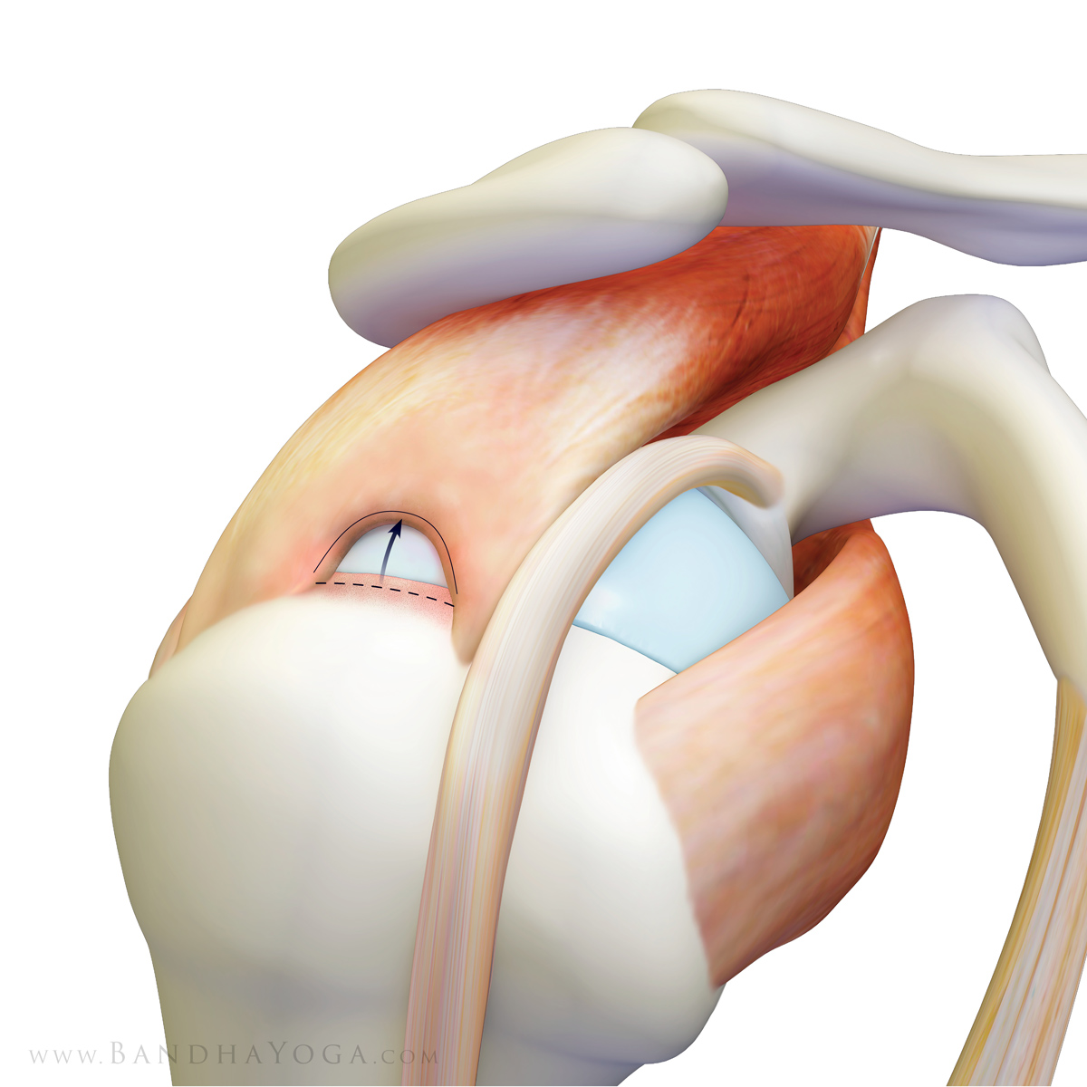
The supraspinatus is the rotator cuff muscle that is most frequently torn. Tears start to become common beyond the age of forty, with an increased incidence in each decade of life. Figure 3 illustrates a supraspinatus rotator cuff tear.


Figure 3: Full thickness tear of the supraspinatus muscle (with the long head of the biceps shown in front of the supraspinatus).


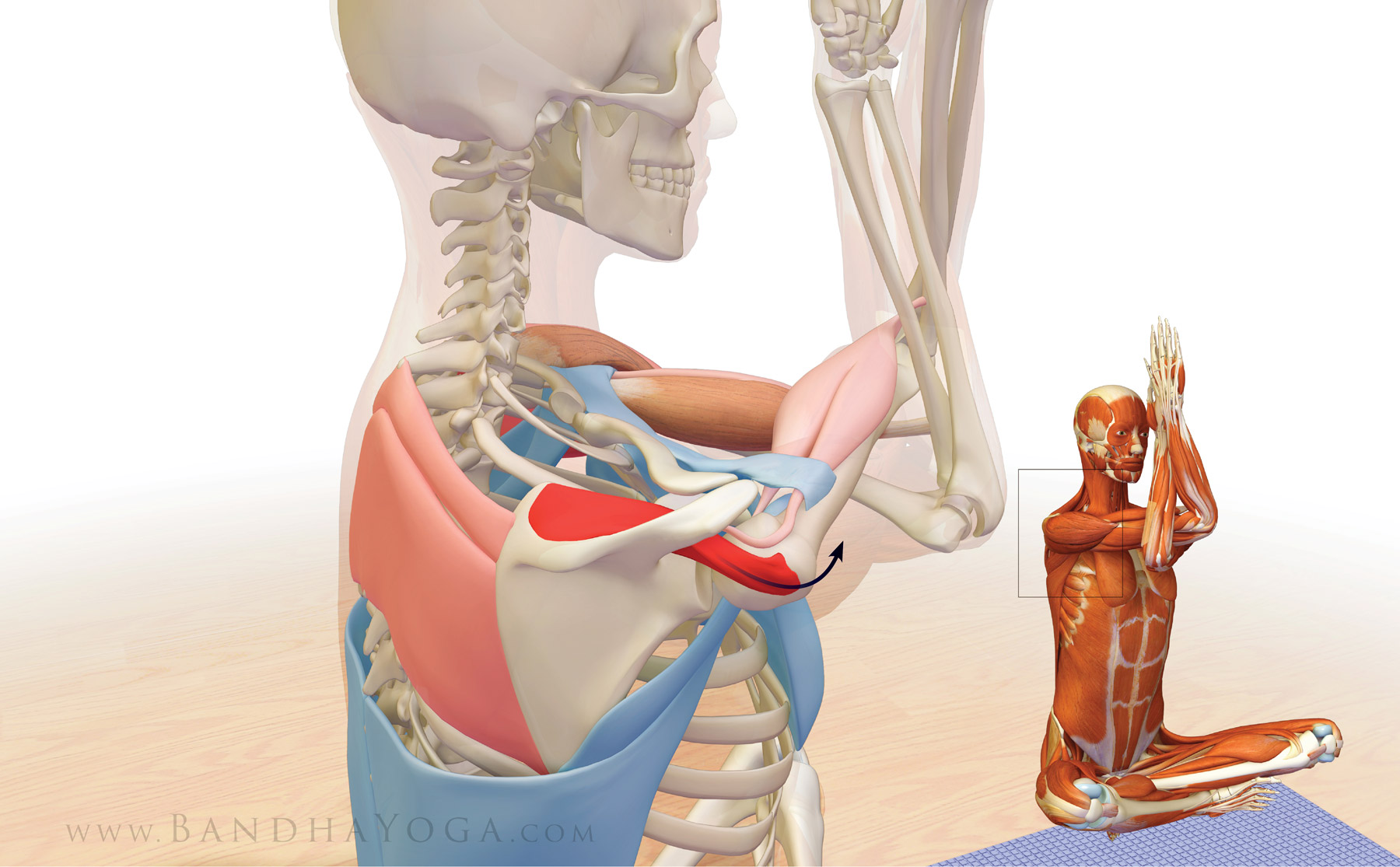
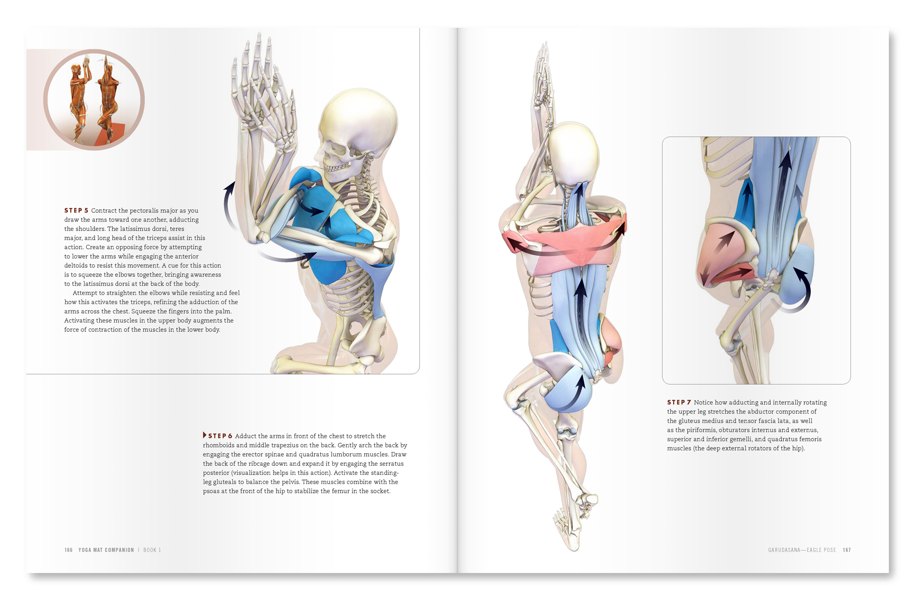
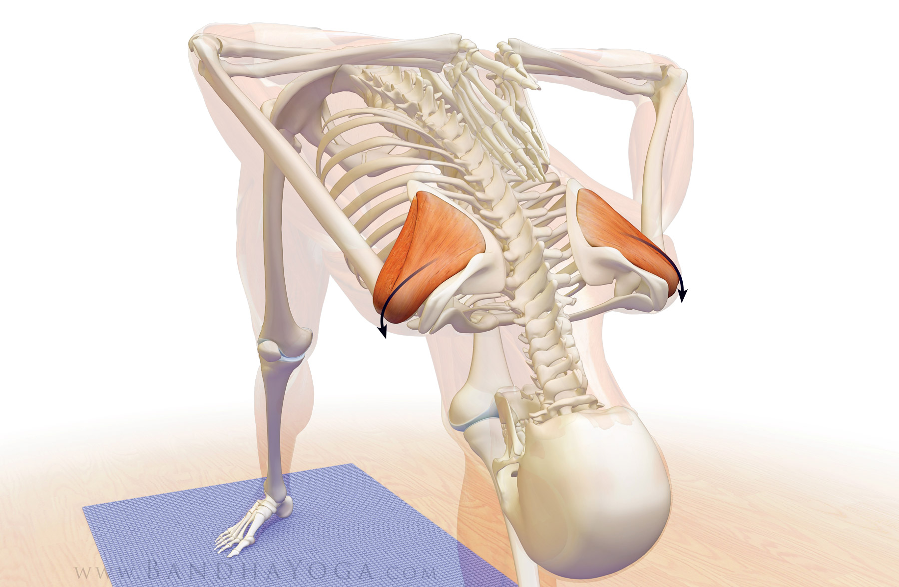
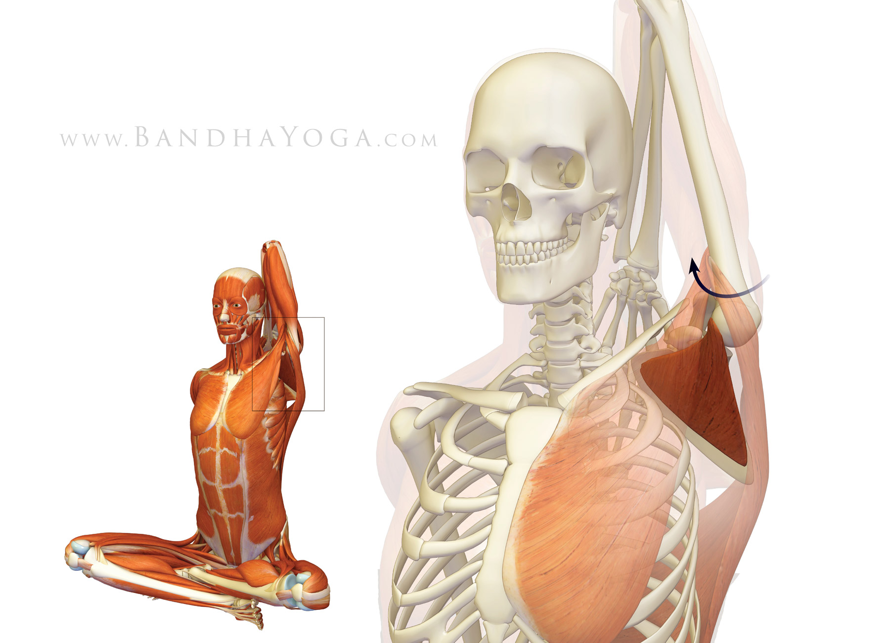
Drawing your arm across the chest (adducting it) stretches the supraspinatus, as well as the capsule of the shoulder and the deltoid muscle. Figure 4 illustrates this action in Garudasana. Note the muscles (colored blue) that contract to stretch the supraspinatus and the muscles that also stretch in this pose (colored red). 


Figure 4: The supraspinatus muscle stretching in Garudasana. The muscles in red are stretching and those in blue are contracting.


Click here to take the rotator cuff quiz to test your knowledge!


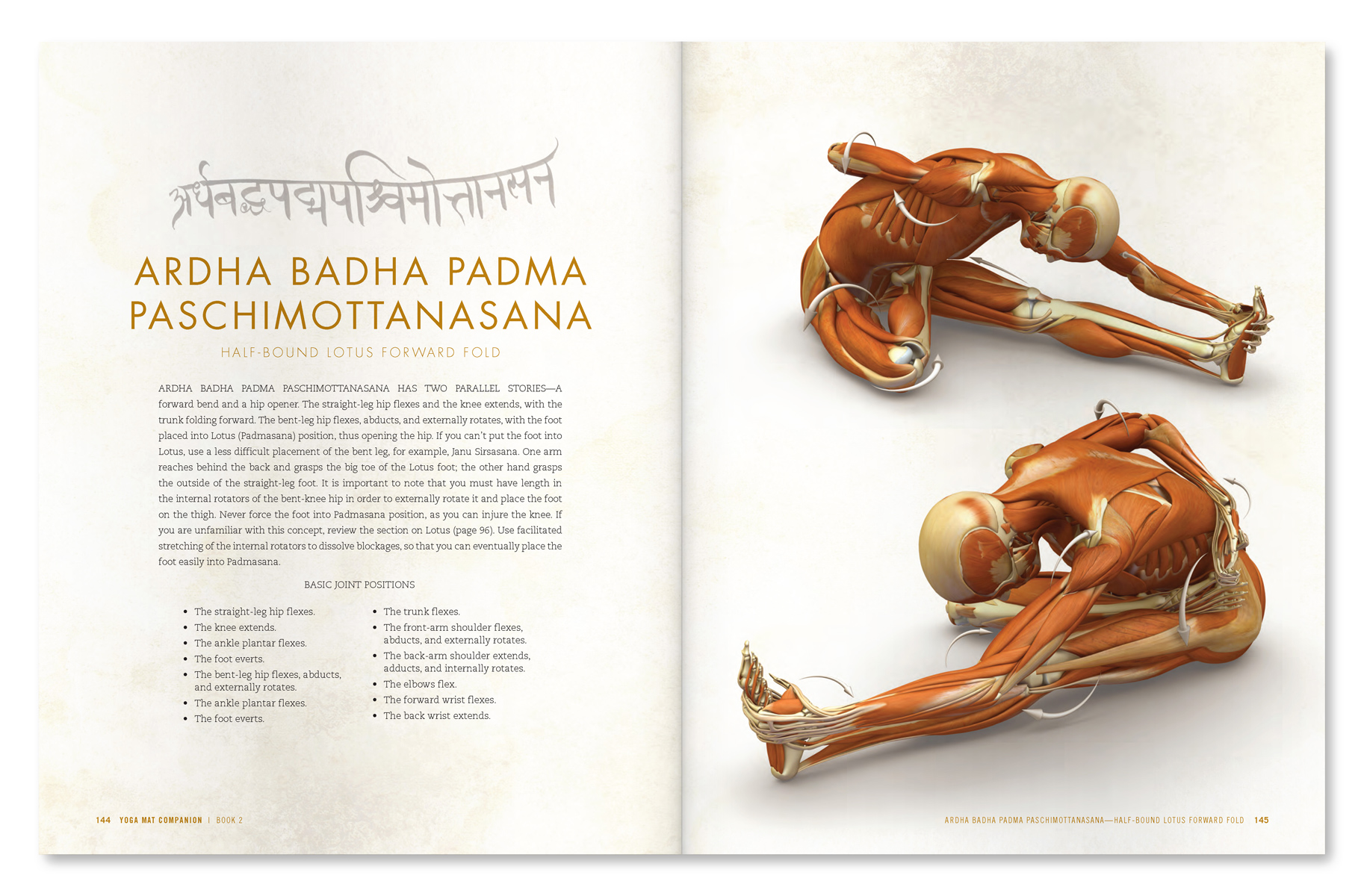
An excerpt from "Yoga Mat Companion 1 - Anatomy for Vinyasa Flow and Standing Poses".


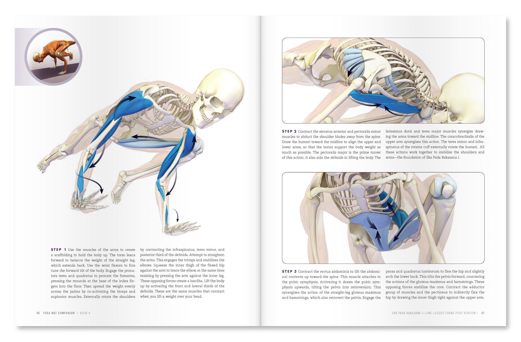
An excerpt from "Yoga Mat Companion 4 - Anatomy for Arm Balances and Inversions".

Thanks for stopping by. Stay tuned for the next post when I'll go over the interaction between the deltoid muscle and the rotator cuff. By the end of this four-post series, you'll have a good understanding of the functional anatomy and biomechanics of the shoulder joint as applied to yoga. Click here to browse through the Bandha Yoga book series on anatomy, biomechanics and physiology for yoga.


All the Best,


Ray Long, MD

Shoulder Biomechanics, Part II: The Infraspinatus & Teres Minor Muscles


Hello Friends,

Welcome to the second of the four-part series on the shoulder joint. Last week I discussed the subscapularis muscle, which is the main shoulder internal rotator. Now we’re on to the antagonist muscles of the subscap, namely, the infraspinatus and teres minor. The infraspinatus arises in a trough below the scapular spine, hence its name (“infra” means below). The teres minor arises back part (dorsum) of the scapula along its outer (lateral) border. The infraspinatus and teres minor insert onto the back part of the greater tuberosity of the humerus, as shown in Figure 1. 

These muscles externally rotate the humerus, with the infraspinatus being the strongest external rotator of the joint. The infraspinatus and teres minor also function to stabilize the humeral head in the socket (glenoid).


Figure 1: The infraspinatus and teres minor muscles of the rotator cuff (the supraspinatus is the faded muscle on top).


The Force Couple

These muscles combine with the subscapularis at the front of the joint to form a “force couple”. In this manner, antagonist muscles (for rotation) become synergists (for stability). Therapy (and surgery) for rotator cuff pathology is directed towards restoring this force couple. Click here to read about concept of antagonist/ synergist combinations for the hip muscles in yoga. Click here for some cues to use this in Dandasana.
Figure 2 illustrates this biomechanical process. This view is looking down on the shoulder with the front of the joint towards the bottom of the page. 


Figure 2: The force couple between the infraspinatus and subscapularis muscles. This view is looking down on the shoulder with the front of the joint towards the bottom of the page.

Poses with the arms in reverse Namaste' stretch the infraspinatus and teres minor, as does Gomukhasana. Those of you who are more flexible may gently press the knife edge of the hand into the back to "load" the external rotators. Folks who are tighter may simply grasp the elbows or hands behind the back. Click here for more details and an animation of Gomukhasana stretching these muscles as well as a not so obvious cue for loading and using PNF for this stretch.

Figure 3: Stretching the infraspinatus and teres minor by internally rotating the shoulders in Parsvottanasana.


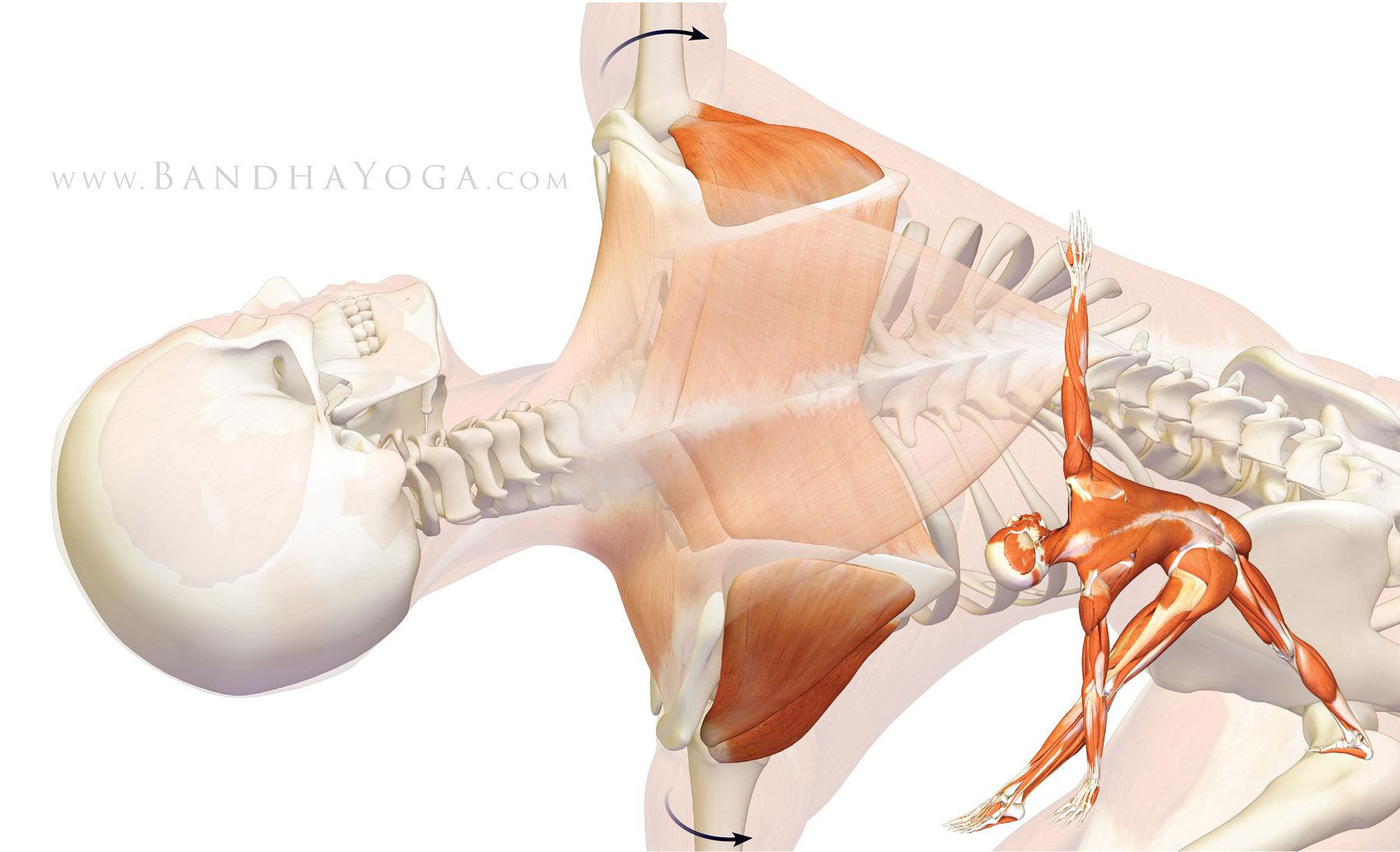
Externally rotating the shoulders in poses like Trikonasana (Triangle) can be used to activate the infraspinatus and teres minor. Figure 4 illustrates this, as well as the myofascial connection between these muscles and the muscles that retract the scapula, namely the trapezius and rhomboids.


Click here to take the rotator cuff quiz and test your knowledge!


An excerpt from "Yoga Mat Companion 4 - Anatomy for Backbends and Inversions".


An excerpt from "Yoga Mat Companion 2 - Anatomy for Hip Openers and Forward Bends".

Thanks for stopping by--I hope you're enjoying learning about biomechanical concepts like the force couple. Stay tuned for the next post when I'll go over the last muscle of the rotator cuff, the supraspinatus. Then I'll finish up with the relationship between the rotator cuff and the deltoids. By the end of this four-post series, you'll have a good understanding of the functional anatomy and biomechanics of the shoulder joint as applied to yoga. Click here to browse through the Bandha Yoga book series on anatomy, biomechanics and physiology for yoga.


All the Best,

Ray Long, MD

Shoulder Biomechanics, Part I: The Subscapularis Muscle

Hello Friends,

This is the first of a four-part series on the shoulder joint, focusing specifically on the rotator cuff and its biomechanical relationship with the deltoid muscle. Let's begin by looking at the muscles that comprise the rotator cuff, starting with the subscapularis. As figure 1 illustrates, the subscapularis occupies the space, or fossa, at the front of the scapula. From there it attaches to the lesser tuberosity, a knob-like structure on the humerus bone at the front of the shoulder. Concentrically contracting the subscapularis muscle (shortening the muscle on contraction) internally rotates the shoulder. The subscap also acts, in conjunction with the infraspinatus muscle, as a stabilizer of the humeral head in the socket (glenoid). We test strength and function of this muscle with the "belly press" test or the "bear hug" test. Tightness in the subscapularis can limit external rotation of the shoulder.


Figure 1: The subscapularis muscle, illustrating the origin on the inside of the scapula and the insertion on the lesser tuberosity of the humerus.


Figure 2 illustrates one of the poses that stretch the subscapularis muscle, namely, Gomukhasana. The upper side humerus externally rotates in this pose, thus stretching the muscle as shown.

Figure 2: This illustrates the effect on the subscapularis muscle of the upper arm in Gomukhasana. External rotation of the humerus stretches the muscle.


Figure 3 illustrates engaging the subscapularis muscle in Ardha Baddha Padma paschimottanasana. Advanced practitioners can attempt to lift the hand off the back to engage the muscle in this pose. This also replicates the "lift off" test, which is used in orthopedics to test the function of the subscap muscle.

Figure 3: This image illustrates contraction of the subscapularis muscle to internally rotate the humerus.


Finally, we have the subscapularis as a stabilizer during a static position in a pose. In Warrior II, attempt to internally rotate the shoulders by imagining pressing the mound at the base of the index fingers down against an object. Resist this by externally rotating the shoulders at the same time. Co-contracting opposing muscles--like the subscap and infraspinatus--stabilizes the head of the humerus in the socket while the deltoid contracts to abduct the humerus. Click here to go into a bit more depth on the subject of stabilizing your shoulders in your Downward dog pose. 


Figure 4: Co-contracting the subscapularis and the infraspinatus stabilizes the humeral head in the socket while the deltoid muscle abducts the humerus.


Click here to take the rotator cuff quiz and test your knowledge!

An excerpt from "Yoga Mat Companion 2 - Anatomy for Hip Openers and Forward Bends".


An excerpt from "Yoga Mat Companion 2 - Anatomy for Hip Openers and Forward Bends".


Thanks for stopping by. Stay tuned for the next post when I'll go over the antagonist muscle for the subscapularis. By the end of this four-post series, you'll have a good understanding of the functional anatomy and biomechanics of the shoulder joint as applied to yoga. Click here to browse through the Bandha Yoga book series on anatomy, biomechanics and physiology for yoga.

All the Best!

Ray Long, MD

Stabilizing Your Shoulders In Downward Dog

Hi Folks,

In our last post, we discussed joint rhythm for the shoulders. In this blog post I want to share some of my recent investigations on the biomechanics of the shoulder joint, with some specific tips for Down Dog. Shoulder pain is one of the problems that comes up in yoga, especially with folks who are doing Vinyasa based practice. The underlying cause of the pain can be multifactorial, but it is frequently related to impingement of the rotator cuff and subsequent inflammation of the cuff tendon (specifically the supraspinatus muscle). Inflammation of the tendon, in turn, affects function of the shoulder. Weakness or instability in the shoulder can then lead to abnormal pressures at the wrist, causing pain there as well. Thus, stabilizing the shoulders has beneficial effects beyond the shoulders. is a complex process involving strengthening the core and then linking the strong core to the shoulders.

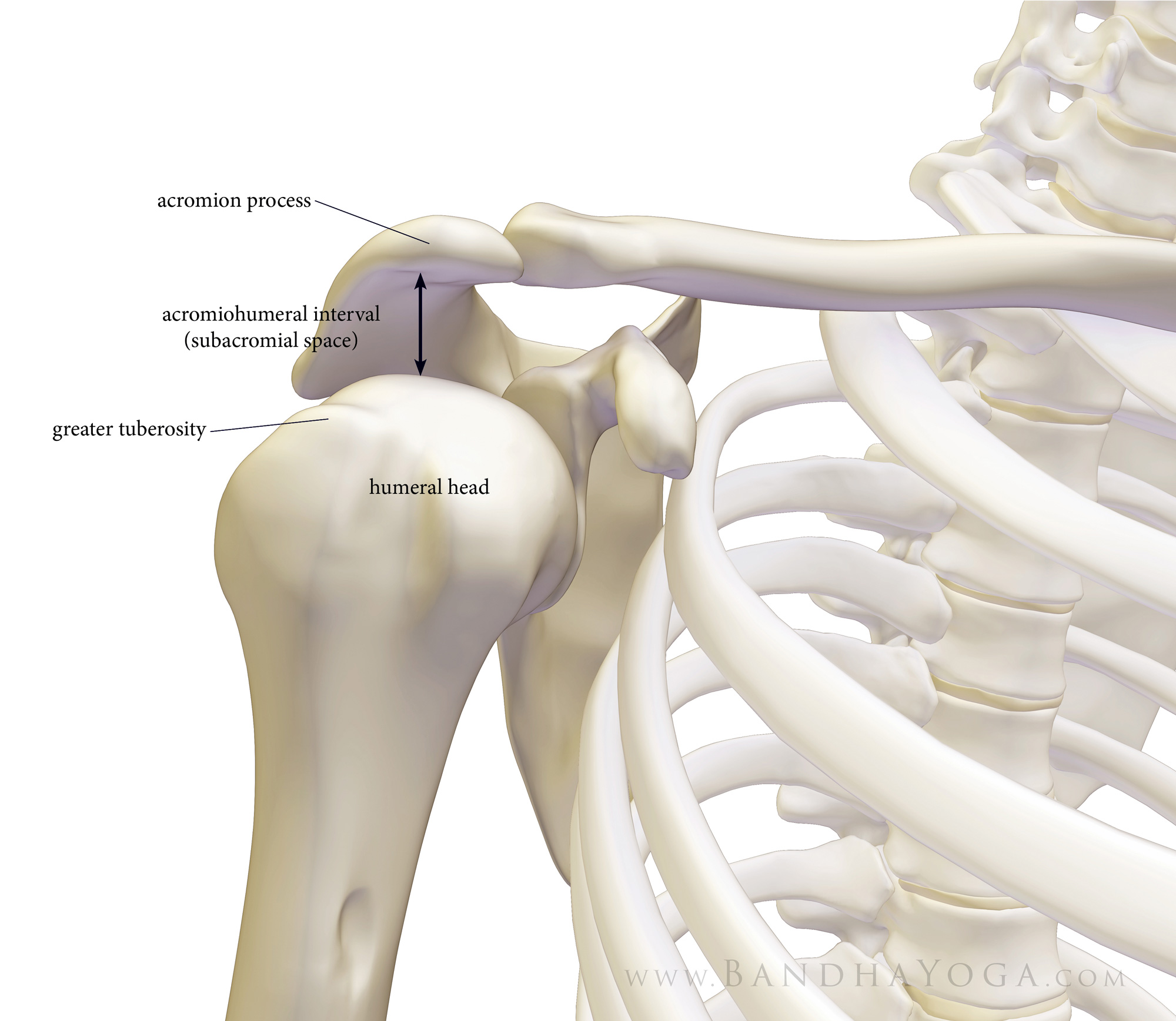
With this in mind, let’s look at one of the key factors in shoulder impingement, namely, the acromio-humeral interval. This refers to the distance between the undersurface of the acromion and the humeral head, as measured using radiology intruments (x-ray, ultrasound, mri). The acromion is a shelf of bone on the scapula, above the spine (seen in Figure 1). It serves as the attachment for the deltoid muscle. The humeral head articulates with the shoulder joint and serves as the attachment for the muscles of the rotator cuff (on the greater and lesser tuberosities). Factors that decrease the space between the acromion and humeral head can lead to inflammation of the cuff tendon due to compression between the two bones.

Figure 1: The acromio-humeral interval. 

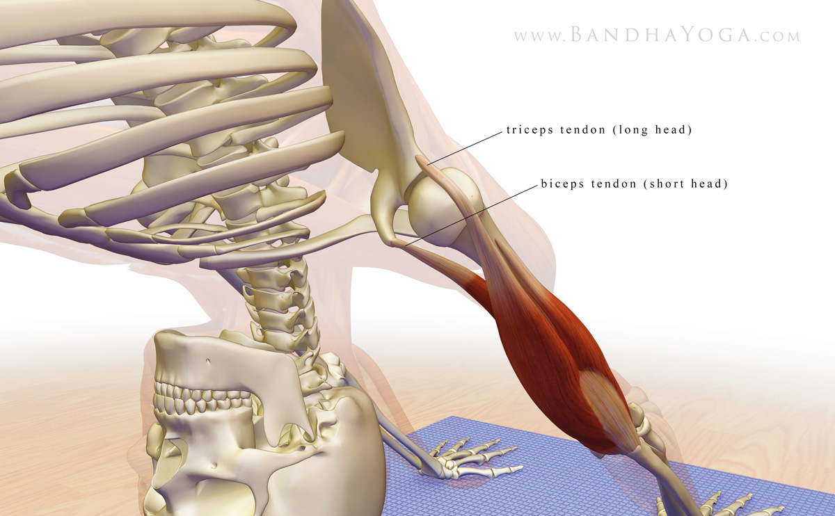
Research has shown that contracting the main adductor muscles of the shoulder serves to increase the acromio-humeral distance. These include the pectoralis major and latissimus dorsi. Co-contracting the biceps and triceps muscles when the arms are overhead can also draw the humerus away from the glenoid, as shown in Figure 2. Finally, externally rotating the shoulder humerus moves the vulnerable area of the supraspinatus tendon away from the area where it would impinge on the acromion (click here to learn more).

Figure 2: The long head of the triceps and short head of the biceps in relation to the gleno-humeral joint with the arms overhead.

Here’s the cue…

Warm up first a bit. Then, take Downward Dog pose. I use three steps for the shoulders. Go slowly and use gentle engagements.

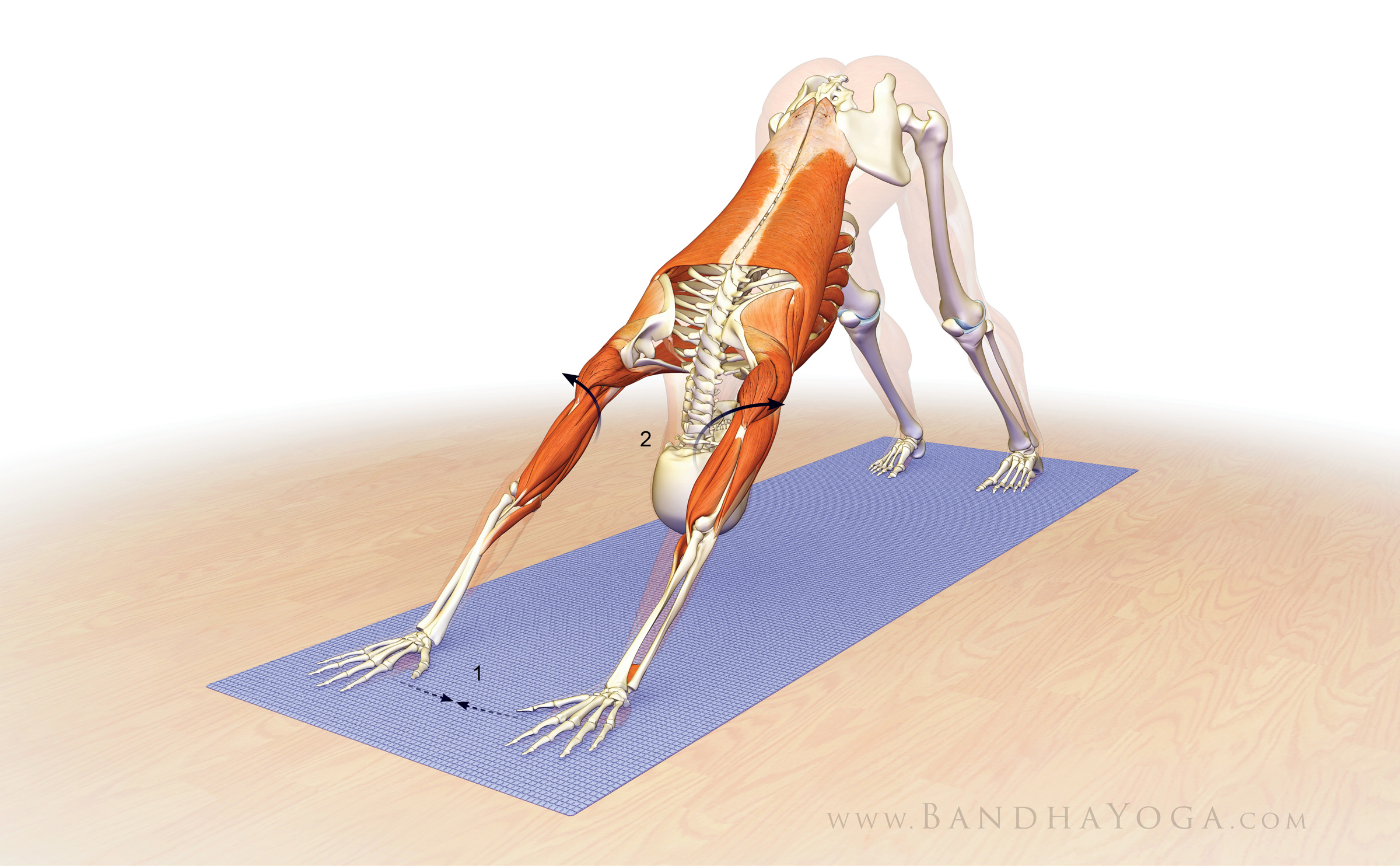
  1. Contract the triceps to straighten your elbows. Then, press the mound at the base of your index fingers into your mat to engage the forearm pronator muscles.
  2. Next, fix your palms into the mat and try to drag the hands towards each other. This engages the adductor muscles of the shoulders as well as the biceps.
  3. Finally, gently roll the shoulders outward. This externally rotates the humerus bone and helps to bring the greater tuberosity away from the undersurface of the acromion.

Figure 3 illustrates the various muscles involved in these cues.

Figure 3: Attempt to drag the hands towards one another. This engages the shoulder adductors. Then externally rotate the shoulders.

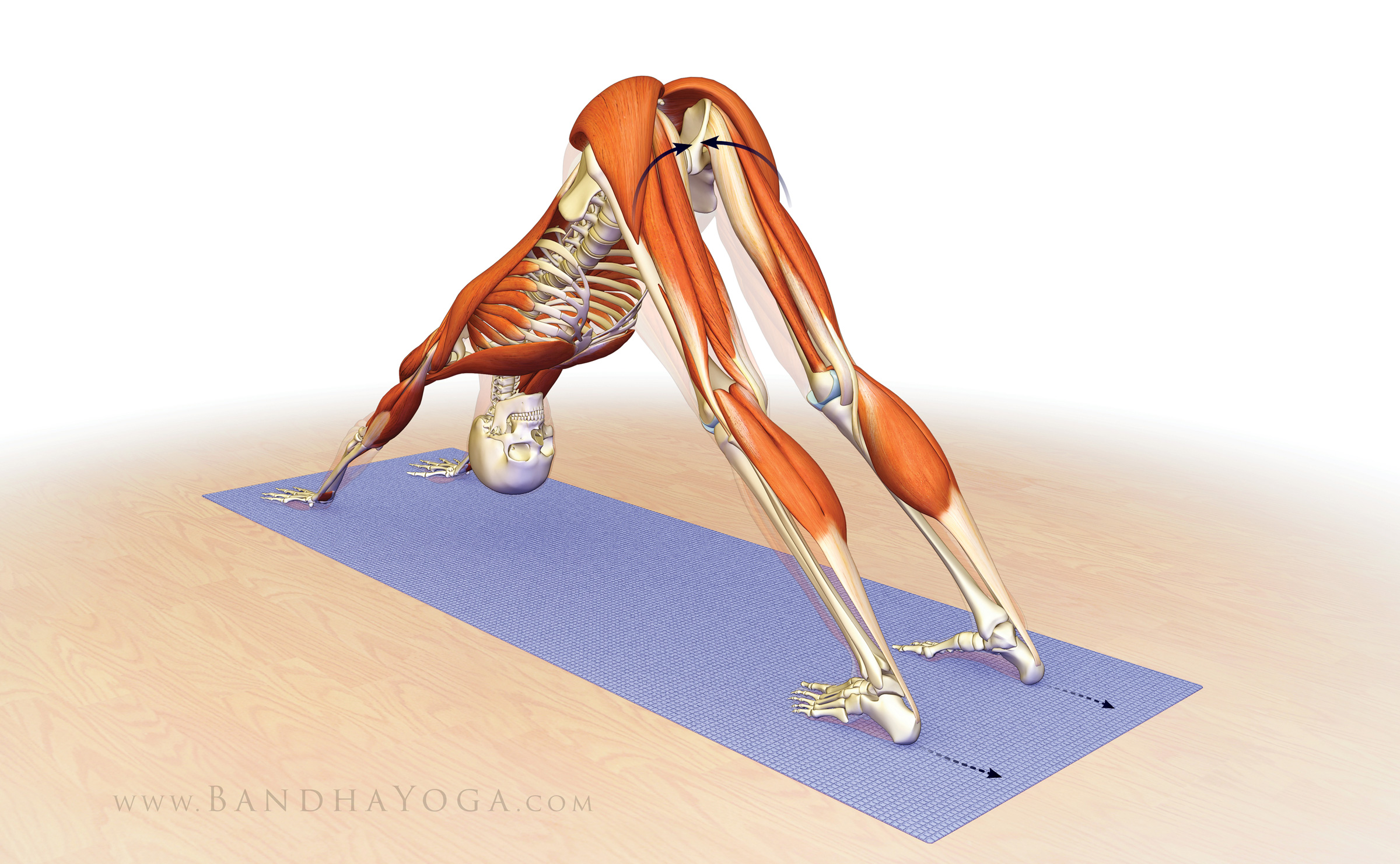
As a final adjustment, I like to link the action of the shoulders to the lower extremities. The cue for this is to engage your lower gluteus max and adductor magnus muscles by drawing in with the upper inner thighs and then attempt to drag your feet away from the hands. Feel how this stabilizes your pose. See Figure 4 for the graphics.

Figure 4: Engage the lower parts of the gluteus maximus and adductor magnus as you attempt to drag the feet away from the hands to stabilize the pose.

Bear in mind that shoulder stability is a complex process. The shoulders are linked to the core; so building a strong core leads to stable shoulders. Stable shoulders help to protect the wrists, and so on. Click here to read more on your core. If you would like to learn more anatomic sequencing to improve your poses, click here to take a tour of The Yoga Mat Companion Series.

An excerpt from "Yoga Mat Companion 1 - Anatomy for Vinyasa Flow and Standing Poses".

An excerpt from "Yoga Mat Companion 4 - Anatomy for Arm Balances and Inversions".


Thanks for stopping by—see you in a couple of weeks for another post on combining anatomy, biomechanics and yoga.

All the Best,

Ray Long, MD


References:


  1. Graichen H1, Bonel H, Stammberger T, Englmeier KH, Reiser M, Eckstein F. Subacromial space width changes during abduction and rotation--a 3-D MR imaging study. Surg Radiol Anat. 1999;21(1):59-64.
  2. Hinterwimmer S1, Von Eisenhart-Rothe R, Siebert M, Putz R, Eckstein F, Vogl T, Graichen H. Influence of adducting and abducting muscle forces on the subacromial space width. Med Sci Sports Exerc. 2003 Dec;35(12):2055-9.

Diaphragmatic (Belly) Breathing

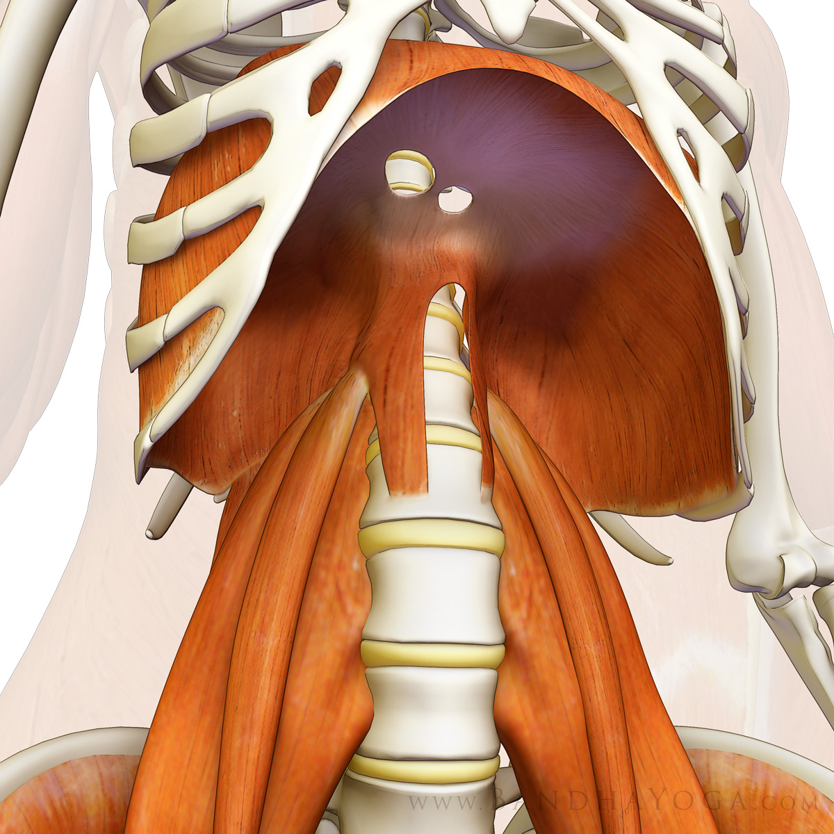
Your thoracic diaphragm is the main engine for breathing, supplemented by the accessory muscles of your chest and abdomen. It is also an important postural muscle with functional connections to your pelvic floor. We'll go over those connections in a future post; in this blog post, let's look at “diaphragmatic” or “belly” breathing.

Figure 1: The thoracic diaphragm (also showing psoas major muscle).

In diaphragmatic breathing, you actively expand the abdomen during inhalation. The abdominal expansion occurs via the diaphragm contracting and pressing down on the abdominal contents. Chest expansion is kept at a minimum in this type of breathing. Exhalation is a relaxed process and occurs through the elastic recoil of the chest wall and lungs.

Regular practice of diaphragmatic breathing draws the mental focus into what is known as the “belly brain”. It has a calming effect on the mind while, at the same time, potentially strengthens the diaphragm. I recommend practicing diaphragmatic breathing for 5-10 minutes per day. We have included a video link below to guide your practice and aid you in visualization of the movement of the diaphragm and abdomen.

Diaphragmatic Breathing Video:


How much does your diaphragm actually move?

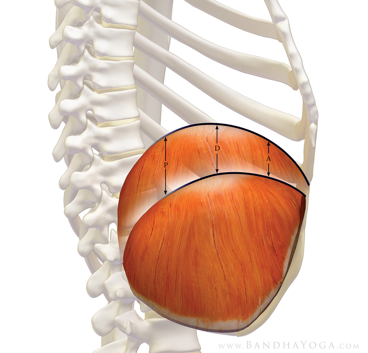
The answer to this question depends on how deep of a breath you take and what part of the diaphragm you are asking about. The diaphragm is a sheet like dome-shaped muscle (when it is relaxed). Upon contraction, it flattens out and presses down on the abdomen. The net result is a negative inspiratory pressure, which draws air into the lungs.

Tidal, or resting breathing results in smaller movements of the diaphragm, while vital capacity breathing (as in a deep diaphragmatic breath) results in much larger movement. This is where you take a complete full inhalation.

The posterior, or back part of the diaphragm exhibits the greatest excursion; the amount of diaphragmatic motion decreases progressively as we come forward. Figure 2 illustrates this. MRI studies (which are considered the most accurate) have quantified diaphragmatic motion during deep breathing, with the posterior region moving an average of 10 cm (about 4 inches) between inhalation and exhalation. This decreases progressively moving forward, with the most anterior portion moving about half that of the posterior. Diaphragmatic motion decreases by about one-third in the sitting position compared to lying on your back. (see reference below)

Figure 2: Thoracic diaphragm (side view): P= posterior; D= dome; A= anterior. Note that the excursion of the posterior diaphragm is greatest.

Does the heart move with your diaphragm when you breathe?

Yes, but not the full excursion of the posterior diaphragm. The pericardium, which is a sac surrounding the heart, has fascial connections to the diaphragm. Accordingly, the heart does move during breathing. Your heart is located more anterior on the left dome of the muscle, and so it moves less than the full excursion of the posterior portions of the diaphragm, but it moves significantly nonetheless. Click here for a video that illustrates diaphragmatic and cardiac movement during breathing (I recommend you start viewing at about the 40 second point, and later at about 4:00 for deeper breathing). This cineradiography video strikingly illustrates this process. (you may also want to mute the sound :)

An excerpt from "Yoga Mat Companion 4 - Anatomy for Arm Balances and Inversions".

An excerpt from "Yoga Mat Companion 1 - Vinyasa Flow and Standing Poses".


Thanks for stopping by. Be sure to have a look at the videos on Youtube. Check back in the next week or so as I have some new info on stretching to share as well.

Learn more about anatomy, biomechanics and physiology for your yoga in “The Key Muscles of Yoga”, “The Key Poses of Yoga” and the Yoga Mat Companion series. Click on any of these books to page through.


All the Best,


Ray Long, MD

and Chris (illustrator/animator)

Kiryu S, Loring SH, Mori Y, Rofsky NM, Hatabu H, Takahashi M.  Quantitative analysis of the velocity and synchronicity of diaphragmatic motion: dynamic MRI in different postures. Magn Reson Imaging. 2006 Dec;24(10):1325-32. 

The Sacroiliac Joint

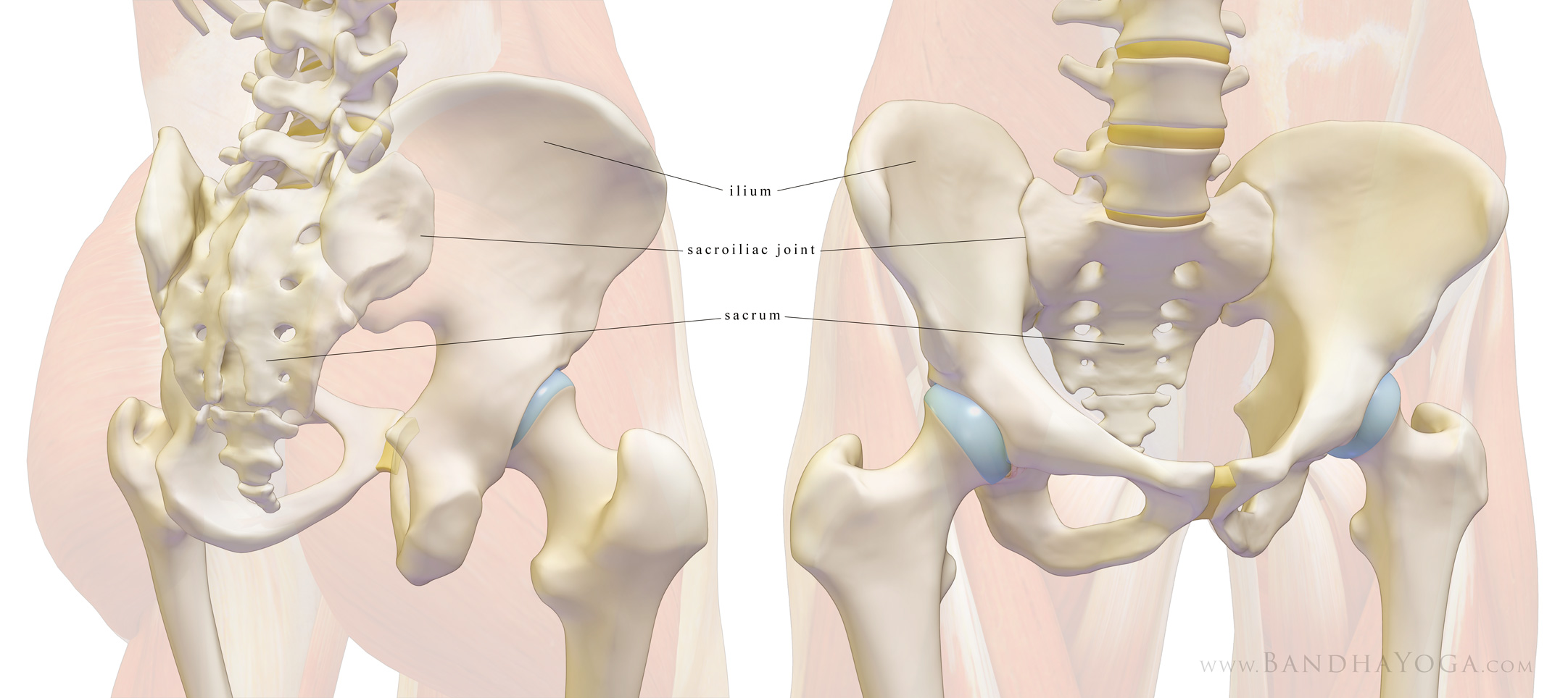
In this blog post we take a look at the fundamental anatomy of the sacroiliac, or SI joint. The SI joint is the articulation between the ilium and the sacrum on each side of the pelvis. As with other joints, it is comprised of the bony stabilizers, the static soft tissue or ligamentous stabilizers, and the dynamic muscular stabilizers. On the surface of the bone is the articular cartilage.

The SI joint depends primarily on the stout ligaments that cross it for stability. The bones also have shallow interdigitations that correspond on each side, thus conferring some bony stability. Finally, there are the muscles (dynamic stability) and fascia—especially the thoracolumbar fascia.

Figure 1 illustrates the bones that comprise the SI joint.

Figure 1: The bones of the sacroiliac joint.


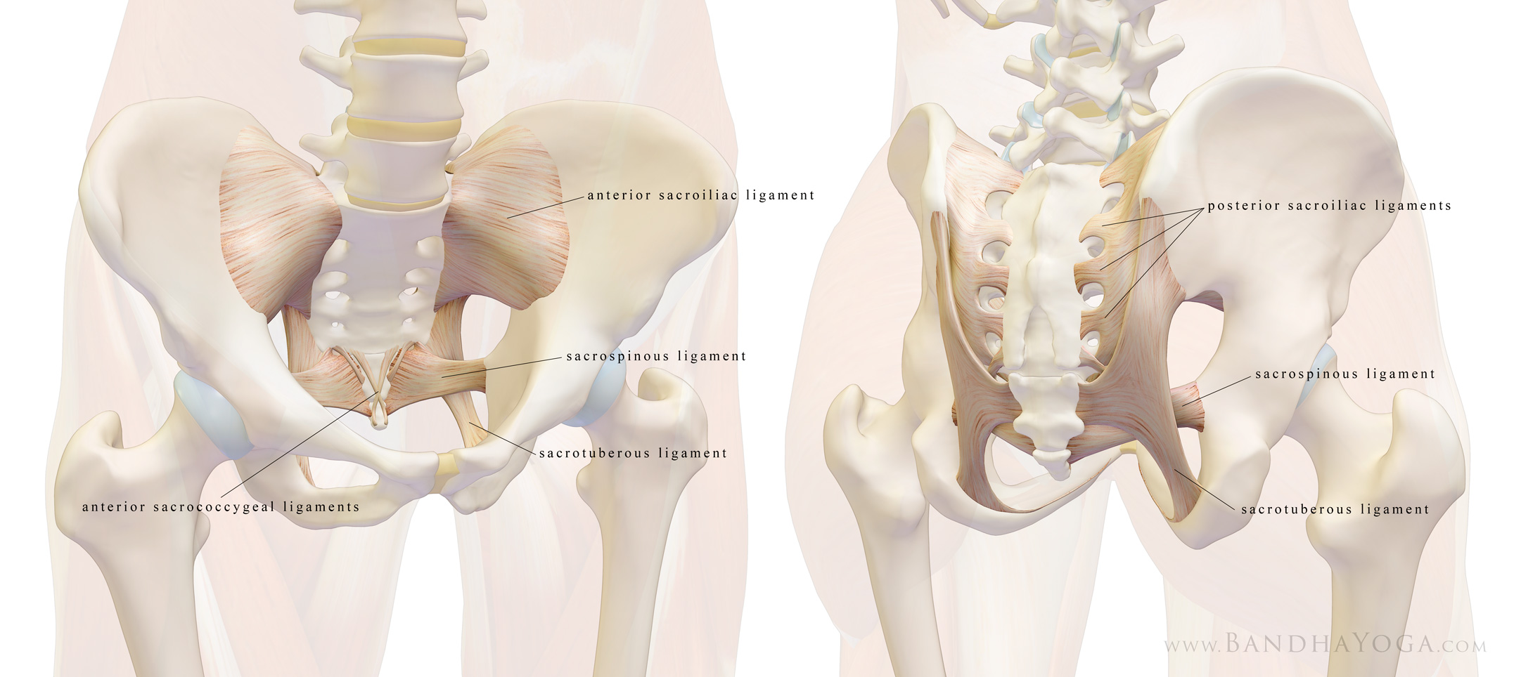
Figure 2 illustrates the stout ligamentous stabilizers of the joint. These include:
  • The anterior (front) and posterior (back) sacroiliac ligaments running from the sacrum to the ilium;
  • The sacrotuberous ligaments running from the sacrum to the ischial tuberosity;
  • The sacrospinous ligaments running from the sacrum to the posterior iliac spine;


Figure 2: The ligaments of the sacroiliac joint.


Movement is very limited for this joint, but includes nutation or anterior tilt (flexion) of the sacrum between the ilia, counter-nutation or posterior tilt (extension) and small movements of the ilia themselves. The stable SI joint thus functions for shock absorption and transfer of torque during ambulation.

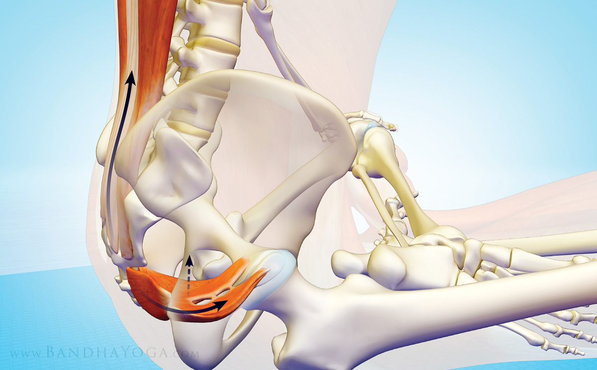
Muscles and fascia also confer stability to the joint. Figure 3 illustrates the relationship between the erector spinae muscles of the back and the muscles of the pelvic floor. You can see that the erector spinae muscles draw the sacrum into flexion (nutation) and the muscles of the pelvic floor (especially the pubococcygeus) draw the bone into extension (counter-nutation). Simultaneously engaging these muscles creates opposing forces that stabilize the joint.

Figure 3: The interaction between the erector spinae and pelvic floor muscles for stabilizing the SI joint.


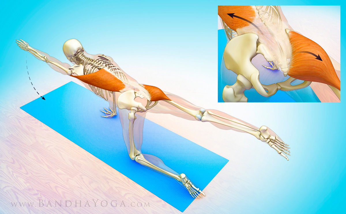
Figure 4 illustrates the relationship of the latissimus dorsi and gluteus maximus muscles on opposite sides of the body. In between is the thoracolumbar fascia. Note how the fibers of these structures run perpendicular to the joint. Thus, working with core exercises such as Bird Dog Pose can help to strengthen the dynamic stabilizers of the SI joint. These muscles, along with the fascia comprise the “posterior oblique subsystem”.

Figure 4: The posterior oblique subsystem for stabilizing the SI joint.


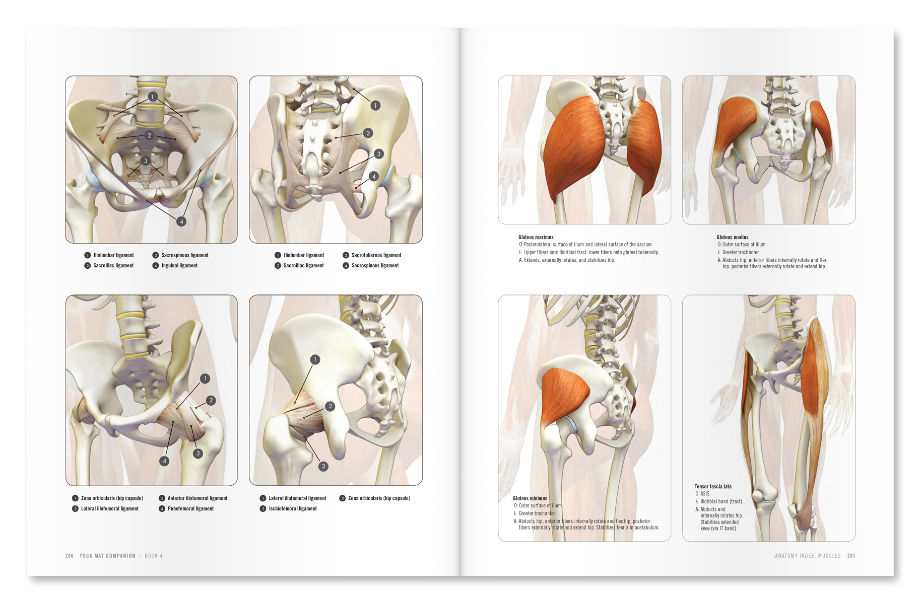
Want to learn more about anatomy and biomechanics for your practice? Click here to check out the Yoga Mat Companion series! Below are some excerpts from these books.

An excerpt from "Yoga Mat Companion 4 - Anatomy for Arm Balances and Inversions".

An excerpt from "Yoga Mat Companion 3 - Anatomy for Backbends and Twists".


Hope you enjoy this overview of the foundational structures of the SI joint. I’d also like to say to our friends in Florida that I'll be teaching in beautiful Tampa/St Pete on the weekend of September 23--25. This is a special intensive in which I will be presenting the most recent research on stretching biomechanics and physiology in a manner that you can apply to your actual practice. Not to be missed! This intensive is the only course I'll be teaching in the Southeast USA this year--hope to see you there! Click here for more information from Suncoast Yoga Teachers Association…


All the Best,

Ray Long, MD

The Pelvic Floor

Dear Friends,

In this blog post I go over the muscles of the pelvic floor. This is an essential structure for support of the pelvic organs; the muscles involved are also engaged in Moola Bandha.

On to the pelvic floor...

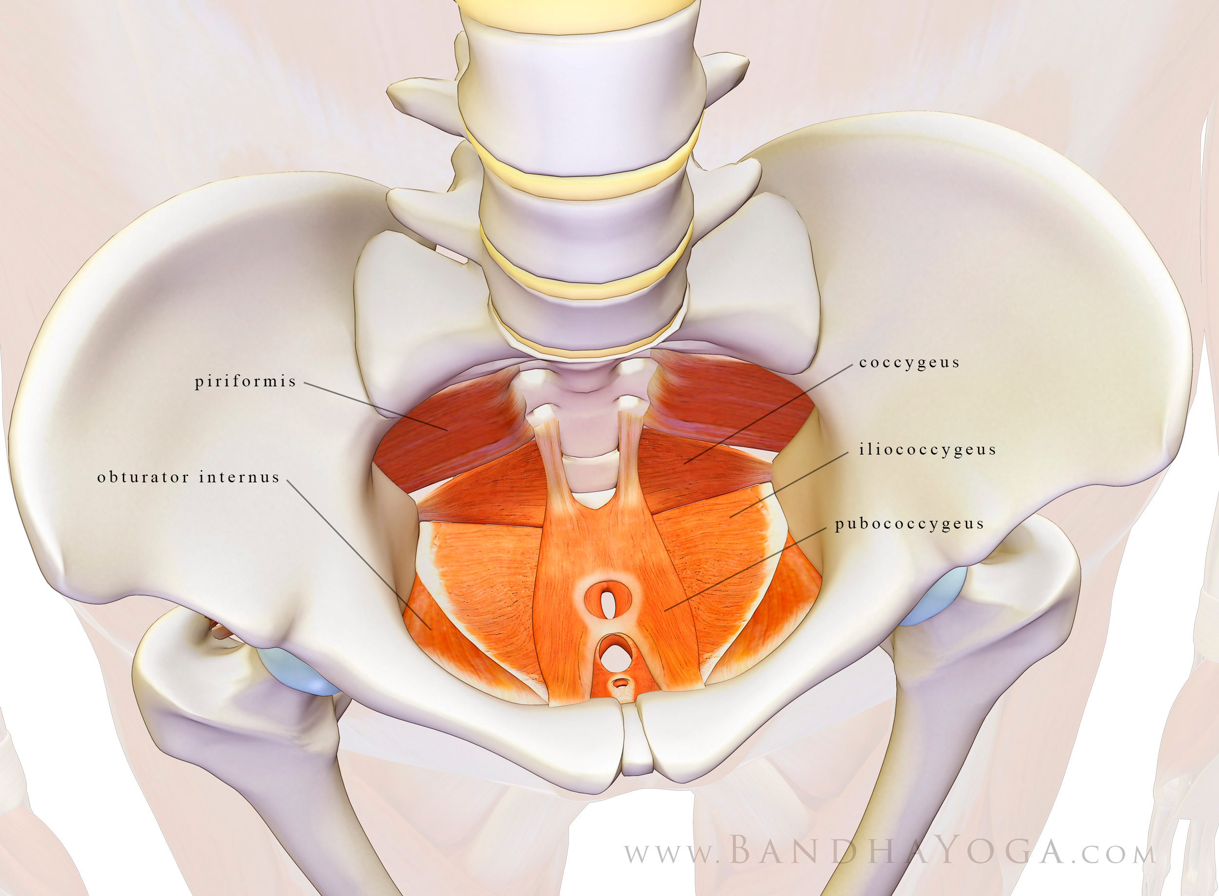
The pelvic floor is comprised of a series of muscles including the piriformis, obturator internus, coccygeus, iliococcygeus, and pubococcygeus. These are illustrated in Figure 1. Other muscles involved include the deep and superficial transverse perineals, the ischiocaveronus and the bulbospongiosus. We illustrate these muscles in Figure 2.

Figure 1: The Pelvic Floor

Figure 2: The Pelvic Floor

Keeping your pubococcygeus strong can help reduce urinary incontinence. All of these muscles provide links to the thoracolumbar fascia, which is linked to the abdominal core. Take a moment to look over these images to get a feel for the attachments of the muscles of the pelvic floor. Kegel exercises and Moola Bandha engage them.


An excerpt from "Yoga Mat Companion 1 - Anatomy for Vinyasa Flow and Standing Poses".


An excerpt from "Yoga Mat Companion 3 - Anatomy for Backbends and Twists".

I’ll have more on this next week—just wanted to give an intro to the structure and let you know about the hacking issue. We appreciate all of your support.

All the Best,

Ray and Chris


Stretching, Aging and Your Down Dog

In this post I discuss how aging affects your body and some recent evidence from the scientific literature on how stretching--and Downward Dog--can help.

First, let’s take a look at reciprocal inhibition…

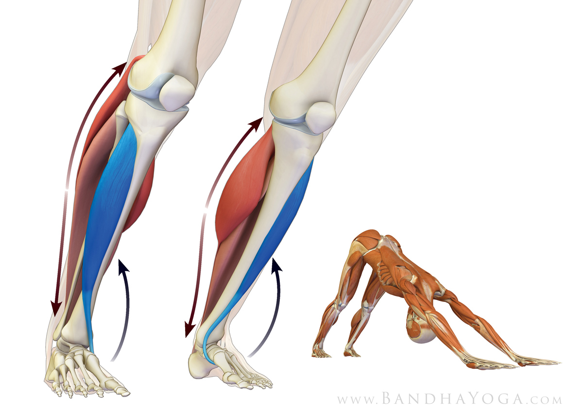
Reciprocal inhibition is a biomechanical and physiological process whereby when we contract a muscle on one side of a joint, the muscle on the other side is inhibited from contracting. This takes place to varying degrees throughout the range of motion of a joint and enables us to do things like walking. For example, when you contract your tibialis anterior to dorsiflex your foot while walking, you automatically inhibit the calf muscles. This helps to prevent catching your toes.

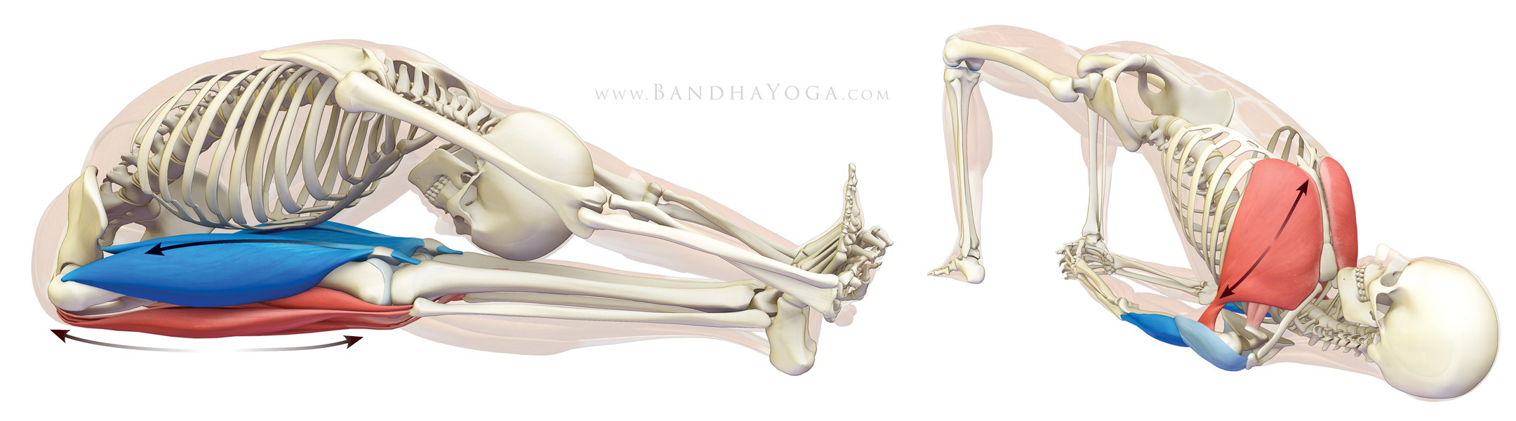
Reciprocal inhibition is controlled and regulated at the level of the spinal cord level and also the brain. Of course, as we reach the ends of range of motion of a joint, a process known as co-contraction kicks in to stabilize the articulation, thus modulating the inhibitory effect of the contracting muscle on its antagonist. In other words, we are not Gumby. (Most folks know this implicitly, but I point it out here to correct some confusing info from recent blogs on the subject of stretching and yoga). Figure 1 illustrates the agonist/antagonist pairs for the forward bend Paschimottanasana and the backbend Setu bandhasana.

Figure 1: Agonist/Antagonist pairs in Paschimottanasana and active Setu bandhasana.

What can happen as we age…

Scientific studies have demonstrated that reciprocal inhibition diminishes as we age (and also in certain disease processes). This is particularly important for activities such as walking. For example, if the tibialis anterior does not efficiently inhibit the gastroc/soleus muscles of the calf, the person tends to catch their toes, stumble and fall. If that weren't bad enough, our bones also weaken with age, so falls can lead to fractures--especially of the hip. This puts the person in a hospital under the care of an orthopedic surgeon as so on…(a story I know all too well). So, methods or techniques that improve reciprocal inhibition could potentially benefit us as we age.

How stretching can help…

Recent scientific evidence has shown that stretching can increase reciprocal inhibition between antagonist muscles. For this study, the authors investigated the effect of stretching on reciprocal inhibition between the tibialis anterior and the gastroc/soleus complex. They stated:

“In conclusion, we have found that 3 wk of twice-daily, static plantar flexor stretching resulted in a significant increase in RI, measured in soleus and gastrocnemius during voluntary, tonic dorsiflexion contractions.” (The full article is linked below in the references)

These are some of the same muscles we work with in Downward Dog. Thus, regular practice of this pose may help maintain reciprocal inhibition between the tibialis anterior and the calf muscles. For a tip on how to make this pose more efficient, take a read through “A Tip to Help You Lower Your Heels In Downward Dog." This blog uses active stretching to improve flexibility of the gastroc/soleus muscle complex. I received a lot of positive feedback on it, so give it a go.

Figure 2: The tibialis anterior as agonist and gastroc soleus as antagonist in Down Dog pose.

On Active Stretching…

In active stretching, one improves muscle flexibility by contracting the opposing muscle group while stretching the target muscle. In the case of the hamstrings, this means engaging the quadriceps during the stretch. Shirley Sahrmann, PhD (Professor of Physiotherapy at Washington University School of Medicine) and others have advocated active stretching as a means of increasing muscle flexibility. It improves flexibility of the muscles on one side of the joint while improving strength and function of the muscles on the other side.

A recent article from the medical literature compared active vs. passive stretching of the hamstring muscles. The authors stated:

“Such an active technique is based on reciprocal inhibition between agonistic and antagonistic muscles.”

And concluded:

“Active stretching produced the greater gain in the AKER test, and the gain was almost completely maintained 4 weeks after the end of the training, which was not seen with the passive stretching group. Active stretching was more time efficient compared with the static stretching and needed a lower compliance to produce effects on flexibility.” (AKER=active knee extension range of motion)

This makes sense to me from a yoga perspective as well--keeping in mind that there are many different ways to climb the mountain. Improving muscle flexibility is only one of the functions of asanas. Active stretching, among other benefits, improves your mind/body connection, alignment of your joints and your mental focus. Improved focus in turn helps to identify and correct imbalances. Click here to read more on the subject of muscle imbalances.

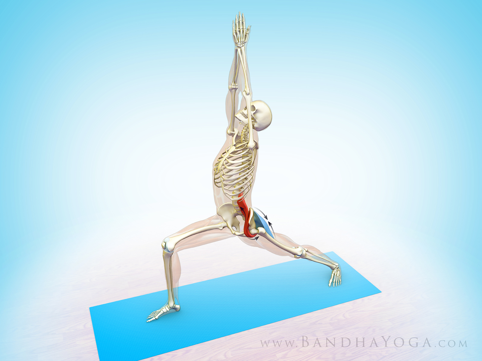
Figure 3: An agonist/antagonist pair in Warrior I (gluteus maximus and illiopsoas).

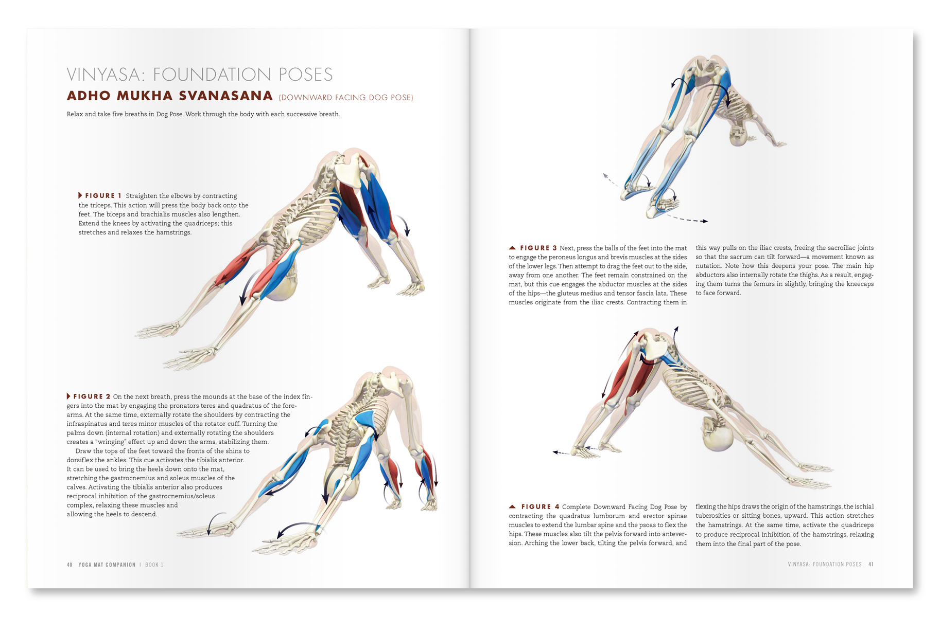
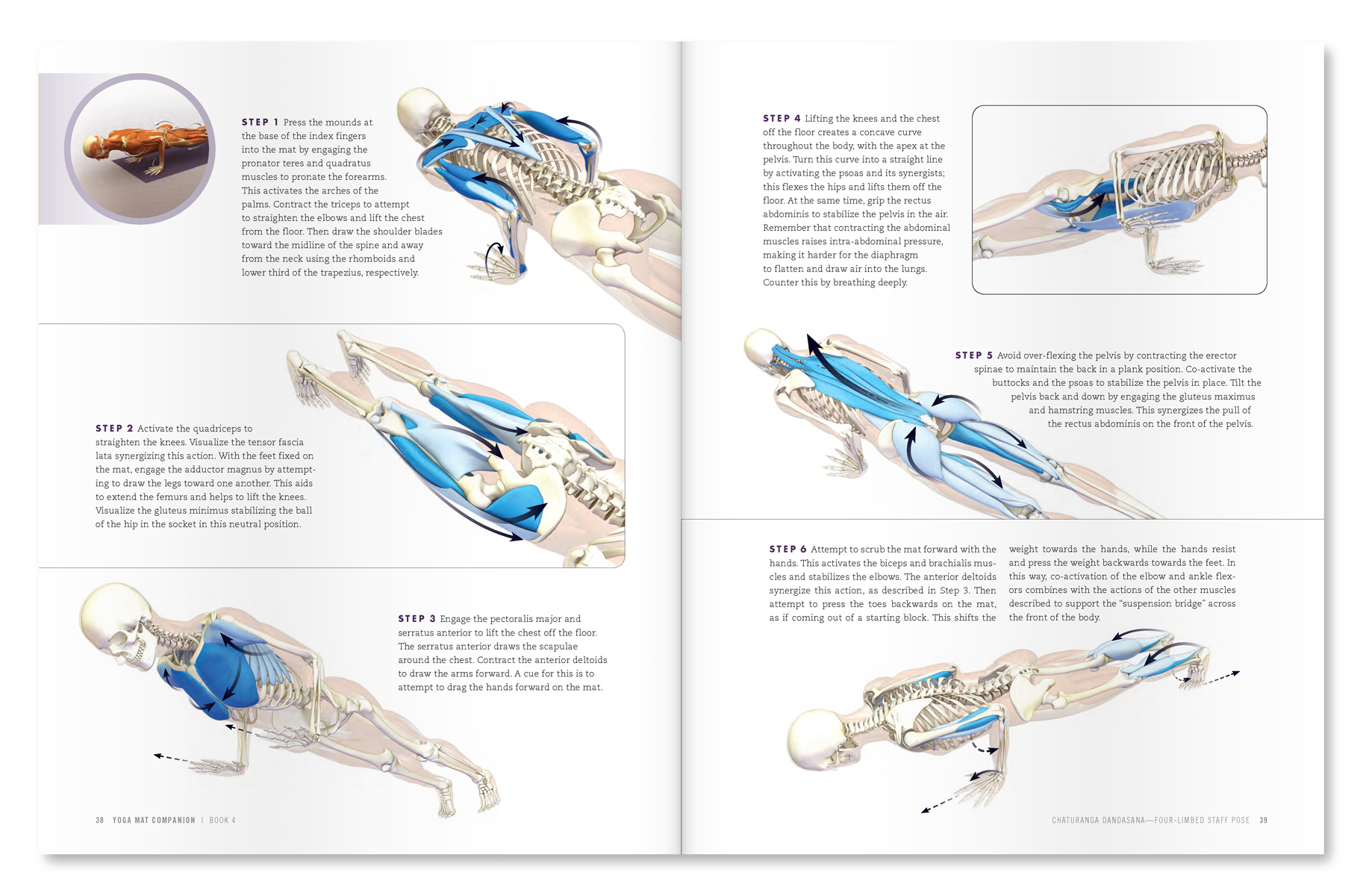
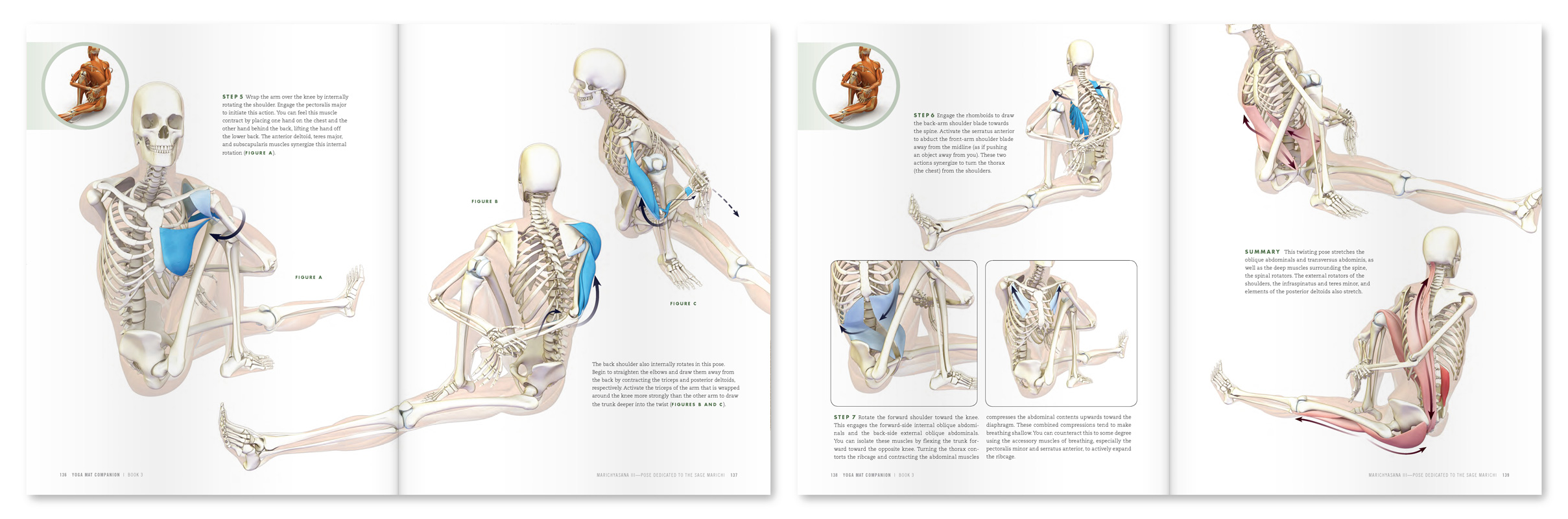
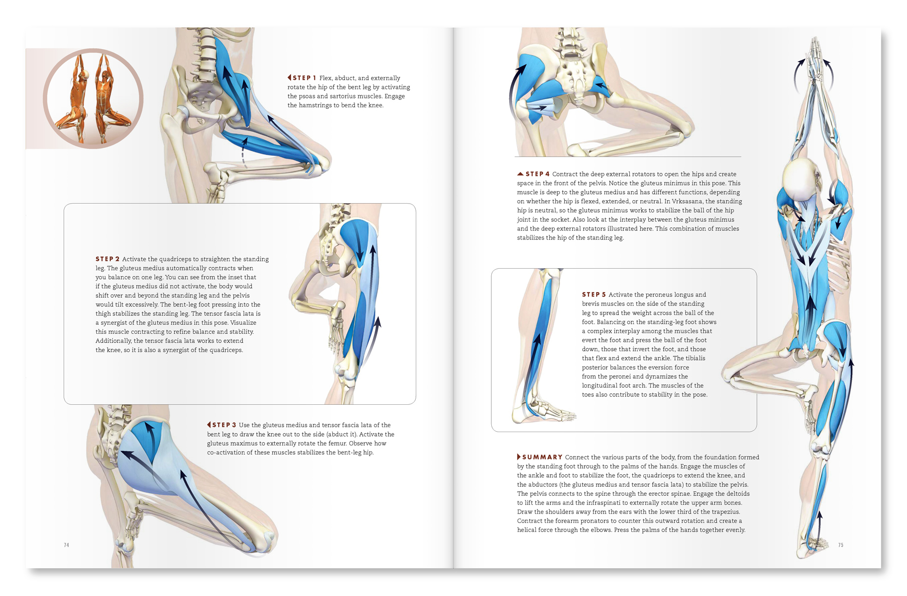
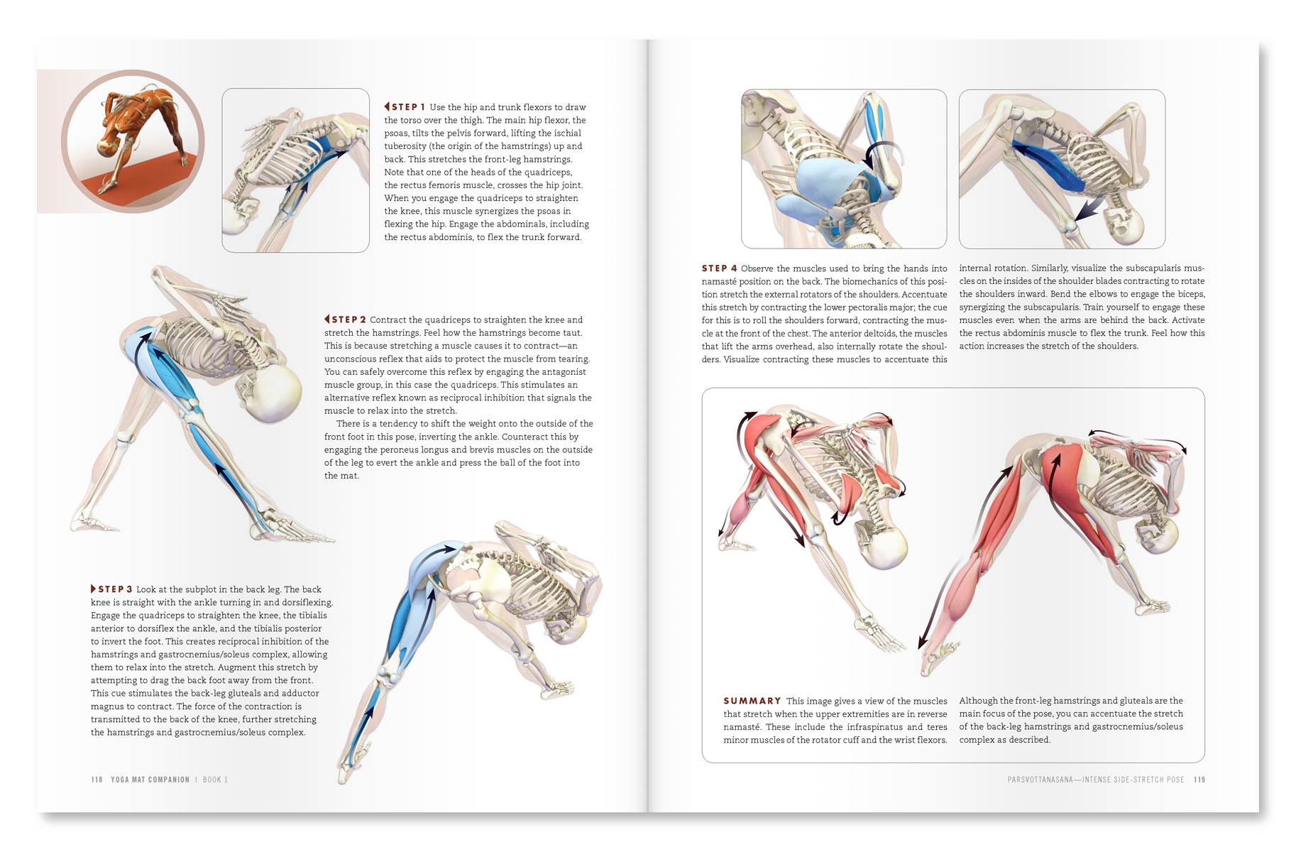
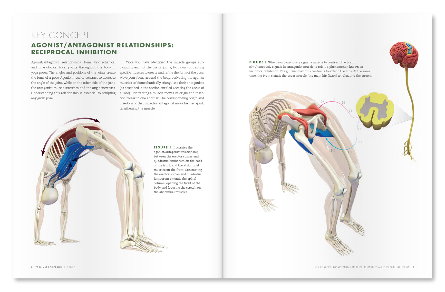
Figure 4 shows a couple of page spreads from our Yoga Mat Companion book series, illustrating a step-wise approach to working with muscle engagement in the poses.

An excerpt from "Yoga Mat Companion 1 - Anatomy for Vinyasa Flow and Standing Poses".

An excerpt from "Yoga Mat Companion 2- Anatomy for Hip Openers and Forward Bends".

NB: Evidence based medicine ranks publications and studies according to “levels of evidence”. Level I studies are considered to be more reliable, and are based on randomized controlled trials. Level VII evidence, on the other hand, is essentially an opinion, with variable reliability depending on the source and their actual expertise. The study on active vs. passive stretching I referenced above was a randomized controlled trial (RCT), and is thus Level I(b) evidence.

Thanks for checking in! Click here for more information on combining modern Western science and your yoga. Hope to see you soon.

All the Best,

Ray Long, MD

References
  1. Lavoie BA, Devanne H, Capaday C. “Differential control of reciprocal inhibition during walking versus postural and voluntary motor tasks in humans.” J Neurophysiol. 1997 Jul;78(1):429-38.
  2. Hortobágyi T, del Olmo MF, Rothwell JC. “Age reduces cortical reciprocal inhibition in humans.” Exp Brain Res. 2006 May;171(3):322-9.
  3. S. Meunier , S. Pol , J. L. Houeto , M. Vidailhet “Abnormal reciprocal inhibition between antagonist muscles in Parkinson's disease” Brain. 2000 May;123 ( Pt 5):1017-26.
  4. A. J. Blazevich , A. D. Kay , C. Waugh , F. Fath , S. Miller , D. Cannavan “Plantarflexor stretch training increases reciprocal inhibition measured during voluntary dorsiflexion” Journal of Neurophysiology Published 1 January 2012 Vol. 107 no. 1, 250-256.
  5. Meroni R, Cerri CG, Lanzarini C, Barindelli G, Morte GD, Gessaga V, Cesana GC, De Vito G. “Comparison of active stretching technique and static stretching technique on hamstring flexibility.” Clin J Sport Med. 2010 Jan;20(1):8-14.
Connect Your Feet to Your Shoulders in Side Forearm Plank Pose

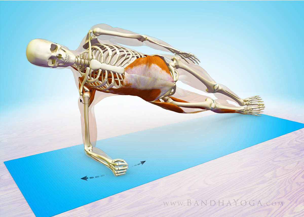
We recently covered the some key poses to strengthen your core, along with biomechanical cues to refine your work in Forearm Plank pose and Bird Dog pose. Side Forearm Plank is another awesome pose to strengthen your core while protecting your wrists. You do this one by placing your forearm on the mat and attempting to drag it towards your feet, while engaging the core muscles on your sides to stabilize the lumbar pelvic complex. Keep your supporting arm (the humerus bone) straight up and down (at a right angle to the floor). This way the passive strength of the bone aids to support your body weight. Click here for more on this concept in Vasisthasana.
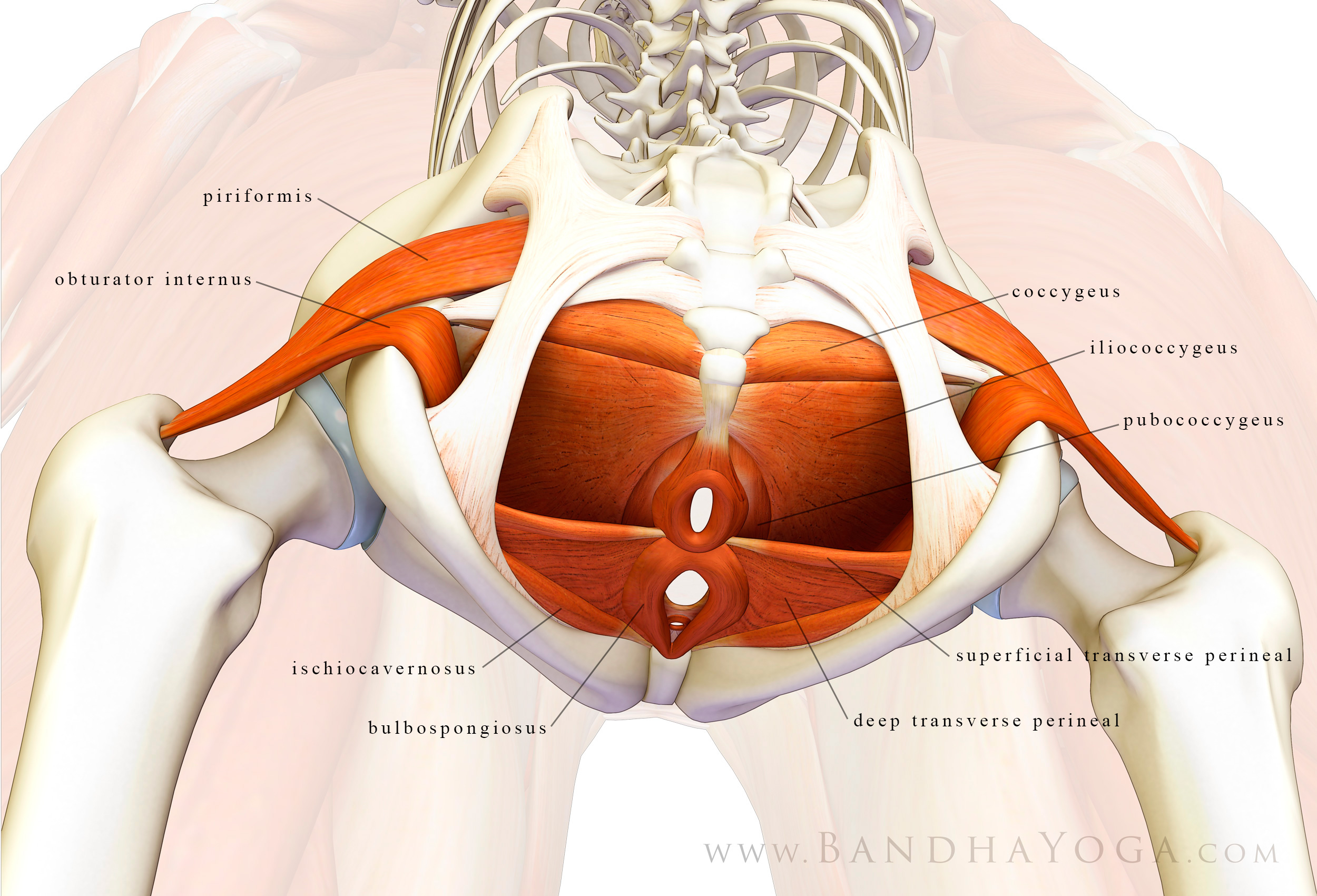
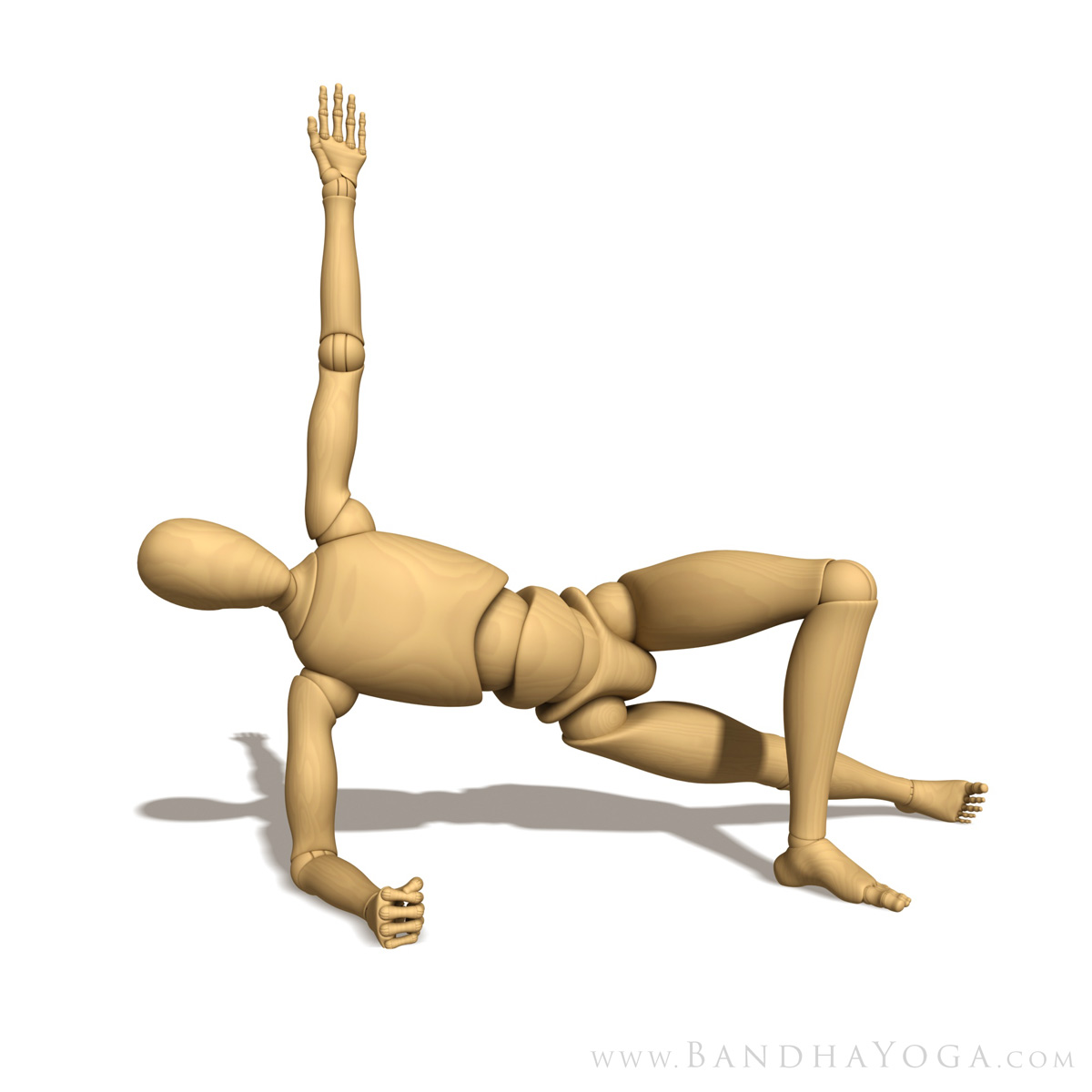
Figure 1: Side Forearm Plank Preparatory Pose

Begin by stabilizing the shoulders. Do this by attempting to externally rotate your forearm on the mat. At the same time, attempt to internally rotate your forearm on the mat as well. It’s a bit like a windshield wiper that’s fixed in place. This cue “co-activates” the infraspinatus and teres minor (external rotation) and the subscapularis (internal rotation) muscles of your rotator cuff. Feel how this stabilizes your shoulder. Folks that are new to this pose can use the preparatory version to work with this cue. Figure 1 shows the prep pose and Figure 2 illustrates the action of the forearms.

Figure 2: This illustrates the cue for co-activating the external and internal shoulder rotators (the infraspinatus, teres minor and subscapularis of the rotator cuff).

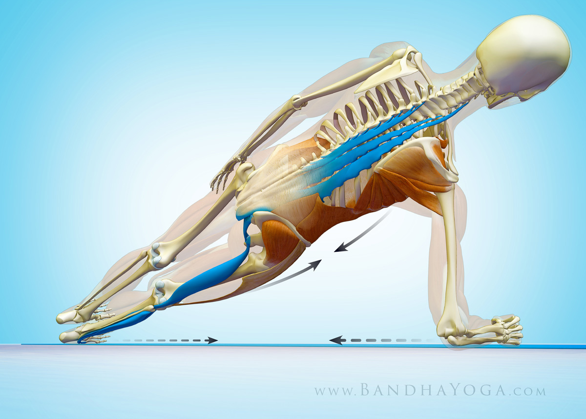
Next, press the edge of your lower side foot into the mat and gently draw it upwards toward the shin to “evert” your foot. These cues activate a series of muscles—including the “lateral subsystem”--to connect your shoulders and legs to your core. Figure 3 shows the cue for attempting to drag the forearm and the feet towards each other (while engaging the side abs).

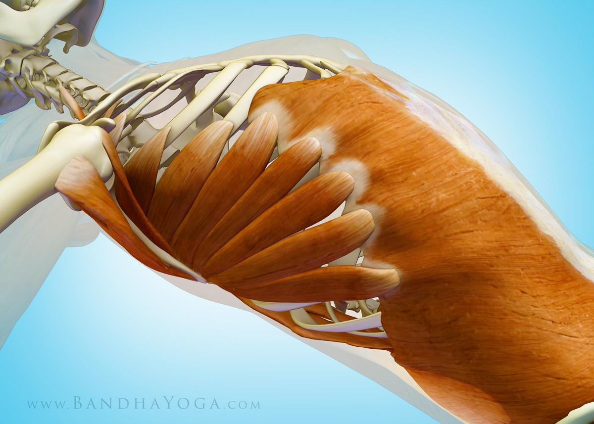
Now let’s check out the myofascial connections in side forearm plank. When you press the side of your foot into the mat, you activate the peroneus muscles as well as the abductor muscles up at your hip (the TFL and gluteus medius). These muscles have a fascial connection to your abs, specifically the external oblique (which attaches to the rim of the pelvis). The external oblique connects to your shoulders via the serratus anterior muscle. The serratus anterior is a scapular stabilizer that works in concert with the rotator cuff. So the whole operation helps to integrate your feet, legs, pelvis and lumbar--all the way up to the shoulders.

Figure 3: This illustrates the cue of everting the lower foot and dragging the elbow towards it. It also shows the deep longitudinal subsystem.

So let’s talk about the deep longitudinal subsystem…

Your deep longitudinal subsystem is made up of the peroneus longus muscle (on the outside of your lower leg), the biceps femoris of your hamstrings and your sacrotuberous ligament (up in the pelvis), the thoracolumbar fascia and the erector spinae muscles (in your back). The biceps femoris creates a link between the lower extremities and the trunk via the sacrotuberous ligament. This ligament helps to transmit force across your sacrum, and, via the thoracolumbar fascia on up the trunk to your deep back muscles. Check Figure 3 for a color coded illustration of this connection. Click here to see this connection in the lower legs in Reverse Pigeon Pose.

This subsystem is part of the global movement system and is thought to be important in force transmission between your trunk and the ground—as in walking. We’ll have more posts on the other subsystems and how to work with them in yoga soon. Click here to see how the abductor muscles of the hip work in your poses. Click here to learn more about the thoracolumbar fascia and its importance in yoga.

Figure 4 shows the myofascial connection between the external oblique muscle (of the abs) and the serratus anterior of the shoulder girdle.


Figure 4: This illustrates the myofascial connection between the external oblique muscle of the abdomen and the serratus anterior of the shoulder girdle.

An excerpt from "Yoga Mat Companion 4 - Anatomy for Arm Balances and Inversions".

An excerpt from "Yoga Mat Companion 4 - Anatomy for Arm Balances and Inversions".

Thanks for checking in! Click here to browse through our books. Or by clicking the links on the right. These books have lots of practical cues with key info on anatomic sequencing to integrate into your practice!


Namaste’


Ray Long, MD